One of the most-discussed topics in healthcare in the year 2018 was „Artificial Intelligence” or just “AI” – and will probably also be in 2019. But what is so controversial about integrating new technology into daily work? It seems like there is concern of computers taking over radiologists’ professional – and later on maybe even personal – lives. This is, of course, not true. The AI-Rad Companion1 is not here to take jobs. It is here to support the daily work of radiologists through smart automation.
Despite all the buzz, not much has really come forward and put AI into a clinical routine workflow. Until now. With an intelligent algorithm to analyze chest CT images, Siemens Healthineers is taking the first big step in establishing a new era of clinical post-processing.

What is this AI-Rad Companion capable of?
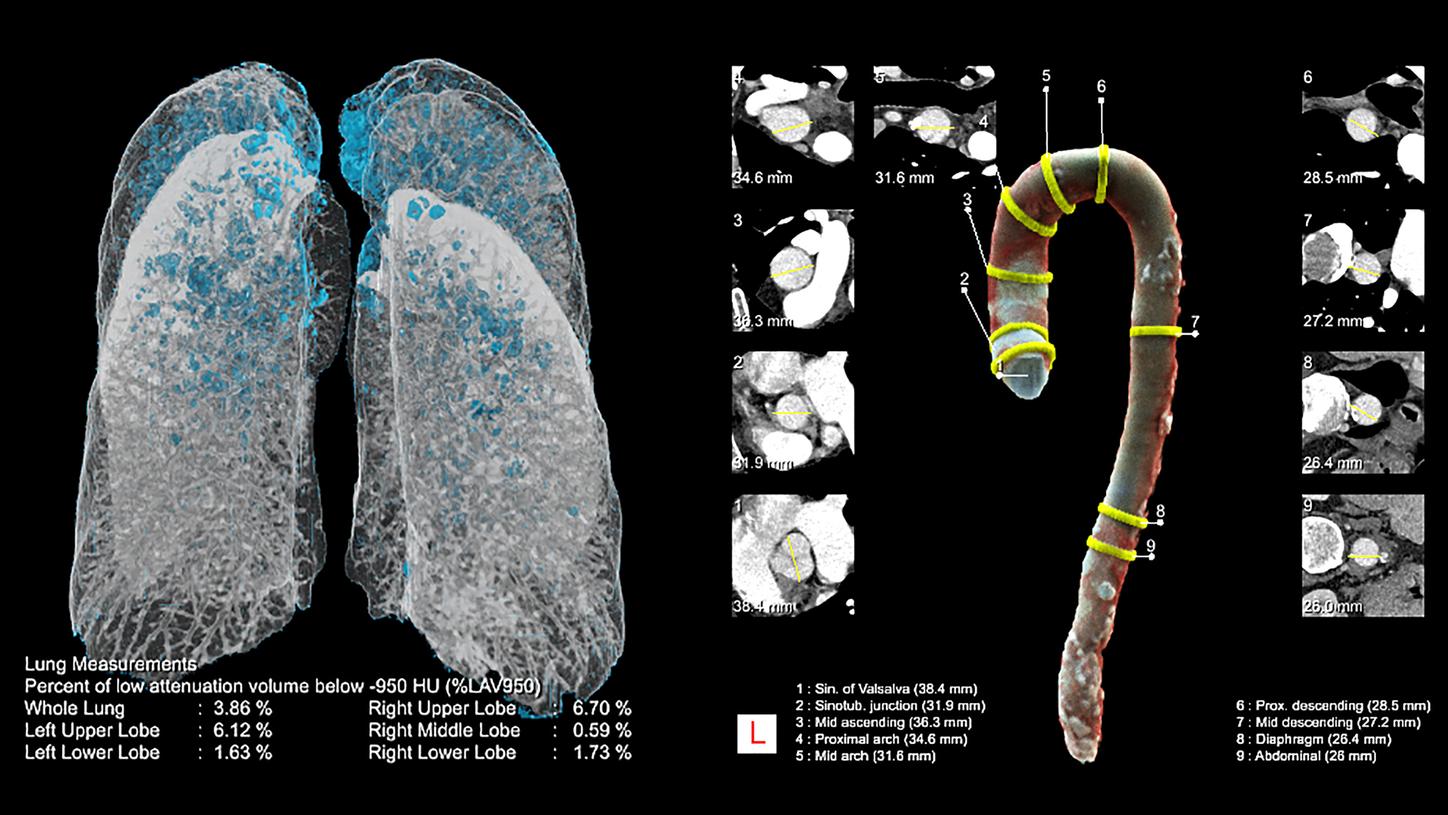
This well-trained algorithm is able to assist radiologists in many ways – for example by measuring the diameter of the thoracic aorta or highlighting suspicious lesions in the lung. And the list goes on: The AI-Rad Companion Chest CT is able to process the following routine steps:
- Highlighting of anatomies and abnormalities
- Quantification of anatomies and abnormalities
- Measuring of anatomies and abnormalities (e.g. diameter of the aorta)
- Reporting of findings in a structured report which is sent directly to PACS
With the AI-Rad Companion radiologists will have the possibility to be supported by an augmented reading aid through workflow optimization and automation.
And what about the humans?
Will radiologists soon be obsolete, then? Absolutely not! The companion will be able to process routine reading and measuring tasks, without inadvertently taking over full control because the level of automation when it comes to sending the datasets to the PACS is configurable. Depending on the preferred level of user-interaction, an additional confirmation dialogue lets the user decide whether or not particular results are sent to the PACS.
Besides that, users might still need to adapt to new tasks and workflows when implementing the AI-Rad Companion into their clinical routine. As always, history tends to repeat itself: With every industrial revolution and every new machine, people needed to put their in-depth knowledge about their craft to a new use. Yet this development is far from being a threat – it’s a chance.
A crucial prerequisite for advancing the implementation of AI and fully exploiting its benefits is therefore the availability of easy-to-use, comprehensive solutions for clinical routine. This applies in particular to the large number of radiologists who generally welcome AI but do not see themselves as tech pioneers. “The early-majority customers expect total solutions for a given business or clinical problem, rather than discrete products and technologies. These solutions must seamlessly integrate into their existing infrastructure,” underscores a current market assessment (Harris 2018).
With the AI-Rad Companion Chest CT, hospitals can rely on their existing IT infrastructure with Siemens Healthineers and therefore a smooth integration into their daily workflow. Although an on-site solution is under active development, this first iteration of the AI-Rad Companion for chest CT is running on the teamplay cloud2. Using this infrastructure, we are following the existing and proven strategy of making data transparent and accessible throughout the hospital or institution.
What about the safety of patient data?
With a heavy focus on privacy and security, patient data is stored with this solution only encrypted and on regional servers according to local regulatory requirements. There is a huge ongoing effort to provide a stable, secure, and reliable technical environment for what is hopefully soon going to be a full suite of clinical AI image-reading algorithms.
More data-driven fields like genetics have already shown that AI could easily become another regular tool in the kit. By fully harnessing its potential, radiologists may potentially gain the freedom to invest their time in better patient care by letting AI do the routine steps and them concentrating on the most important: the patient.

