AI-Rad CompanionPodpora při rozhodování u multimodálního zobrazování
AI-Rad Companion, naše skupina řešení rozšířeného pracovního postupu s umělou inteligencí, vám pomůže snížit zátěž základních opakujících se úkolů a zvýšit diagnostickou přesnost při interpretaci lékařských snímků.
Řešení poskytují automatické následné zpracování obrazových datových sad prostřednictvím algoritmů využívajících umělou inteligenci. Automatizace rutinních pracovních postupů s opakujícími se úkoly a velkým objemem případů vám pomůže usnadnit každodenní práci a můžete se soustředit na zásadnější problémy.
Training of algorithms for AI-Rad Companion Chest CT

Training of algorithms for AI-Rad Companion Chest CT

Training of algorithms for AI-Rad Companion Chest CT
Aktuální algoritmy a škálovatelné nabídky
Naše nejmodernější algoritmy vám jako uživateli budou automaticky distribuovány, jakmile budou oficiálně vydány a zpřístupněny. Pokud snímky následně zpracuje AI-Rad Companion, podpoří vaši interpretaci dat tím, že vám automaticky poskytne výsledky své analýzy ke kontrole, potvrzení a případnému zahrnutí do závěrečné zprávy nebo postupu péče – pro zvýšení vaší přesnosti a zajištění vyšší jistoty při diagnostickém rozhodování.
Všechna rozšíření AI-Rad Companion jsou nasazována prostřednictvím platformy teamplay. Tento přístup usnadňuje pravidelné aktualizace i upgrade a usnadňuje integraci nových nabídek do stávajícího IT prostředí.
AI-Rad Companion skupina se rozrůstá

Jak se AI-Rad Companion využívá v praxi
Zákazníci po celém světě používají AI-Rad Companion ve své klinické praxi. Zjistěte, jaké mají pozitivní zkušenosti od doby, kdy začali používat AI-Rad Companion Chest CT ve své každodenní práci.
Customer experiences with AI-Rad Companion Chest CT
Clinical Outcomes
AI-Rad Companion may help you to increase precision and speed up your workflow
We developed the AI-Rad Companion to help you cope with a growing workload. With its deep learning algorithms, AI-Rad Companion automatically highlights abnormalities, segments anatomies, and compares results to reference values.
Discover how AI-Rad Companion supports you as a full-time radiology expert
Brain MR
04The AI-Rad Companion Organs RT automatically contours the organs at risk with the support of deep learning algorithms. It may reduce unwarranted variations with high-quality contours that approach the level of consensus-based contours.
Courtesy
of Leopoldina-Krankenhaus der Stadt Schweinfurt GmbH, Schweinfurt, Germany

The AI-Rad Companion Chest CT detects and highlights lung nodules. After segmentation of the lung nodules, the volume, maximum 2D diameter, maximum 3D diameter and tumor burden are automatically calculated.
Courtesy of Klinikum Nürnberg, Nuremberg, Germany

Cinematic Rendering adds clarity to the location of a detected lung nodule. The visualization makes it easy for the referring physician to get a clear picture of the nodule location within the lung.
Courtesy of Klinikum Nürnberg, Nuremberg, Germany

The AI-Rad Companion Chest CT automatically recognizes coronary calcium, calculates its volume, and creates an enhanced clinical image including quantification of the heart volume. This additional information about a cardiac condition is valuable clinical knowledge to a routine Chest CT study.
Courtesy of Klinikum Nürnberg, Nuremberg, Germany

This 3D overview of the thoracic aorta has been automatically created by the AI-Rad Companion Chest CT. It includes the measurement of relevant diameters, based on medical guidelines and detected anatomical landmarks. This clearly shows how the AI-Rad Companion Chest CT can support the increase of accuracy of your reporting.

The AI-Rad Companion Chest CT has automatically created this measurement of the aorta based on a non-contrast enhanced CT image.
Courtesy of Klinikum Nürnberg, Nuremberg, Germany

On this image you can see how the height of the vertebrae bodies is measured and compared. The AI-Rad Companion Chest CT automatically processes these measurements and marks abnormalities with easy-to-understand color-coding. Deviations are highlighted, while bone density is reported in Hounsfield Units.
Courtesy of Klinikum Nürnberg, Nuremberg, Germany

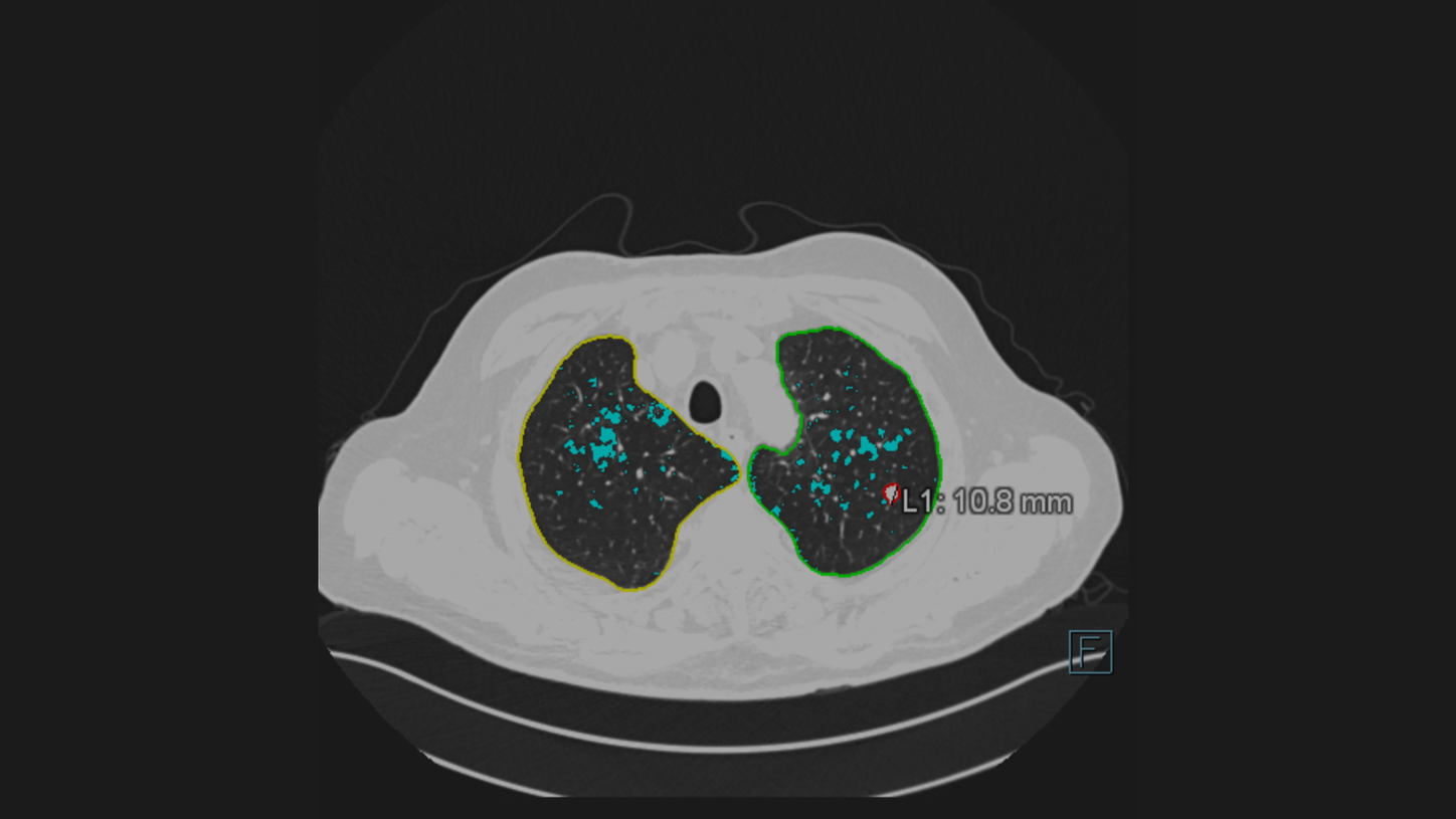
The new functionality Pulmonary Density3 automatically identifies and quantifies hyperdense areas of the lung using non-contrast enhanced chest CT’s. The opacity score helps to assess the percentage of affected lung tissue.

Certain parts of the pulmonary density functionality1 of AI-Rad Companion Chest CT were used in the prototype Siemens Healthineers offered to fight COVID-19, to analyze ground-glass opacities and consolidations. High opacity abnormalities were shown to correlate with lungs of COVID-19 patients.
For this sub mSv CT examination the Tin Filter technology is used.
kV: Sn130 kV, Q. ref mAs: 40 mAs, CTDIvol: 1,38 mGy DLP: 51
Eff. Dose: 0,86 mSv
Courtesy of CHR East Belgium, Verviers, Belgium

A 360° VRT overview visualizes the affected areas.
Courtesy of CHR East Belgium, Verviers, Belgium

Neurology - Artificial intelligence supports clinicians in reading brain images
A new software helps radiologists view, analyze, and evaluate brain images.

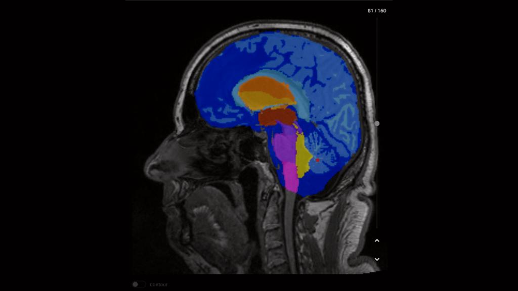
AI-Rad Companion performs an automatic segmentation of the different brain structures , and provides individual volumetric analysis.
Courtesy of CHUV, Lausanne, Switzerland, Case 2aaaa1366

AI-Rad Companion compares the different volumes to a normative database and automatically generates a highlighted deviation map.
Courtesy of CHUV, Lausanne, Switzerland, Case 2aaaa1366

AI-Rad Companion automatically generates a report with all volumetric analyses listed, deviations from the normative database are marked. All results can be transferred to PACS based on the configuration.
Courtesy of Centre d'imagerie diagnostique, Lausanne, Switzerland, Case 2aaaa1366

AI-Rad Companion Prostate MR for biopsy support performs an automated segmentation as well as an automated volume estimation of the prostate. When the PSA value is known, it calculates the PSA density.
Courtesy of Clinica Universidad de Navarra, Spain

The radiologist can manually mark and characterize lesions and other targets and add comments within the AI-Rad Companion Prostate MR for biopsy support. The solution can export for transfer the segmentations and targets found, as well as burnt-in contours, by a DICOM standards based radiotherapy structure set (RTSTRUCT). These images can be exported and used by the urologist to fuse with Ultrasound images as a biopsy guidance.
Courtesy of Clinica Universidad de Navarra, Spain

PA chest X-ray of a patient showing AI-Rad Companion Chest X-ray findings of two lung lesions in the left lung and the consolidation in the right lung.
Courtesy of MVZ Prof. Dr. Uhlenbrock & Partner GmbH, Dortmund, Germany

PA chest X-ray of a patient showing an AI-Rad Companion Chest X-ray lung lesion finding in the right lung and
atelectasis in the left lung
Courtesy of MVZ Prof. Dr. Uhlenbrock & Partner GmbH, Dortmund, Germany

CT scan of the pelvis with AI-Rad Companion Organs RT generated contouring of organs at risk.
Courtesy
of University Hospital Erlangen, Erlangen, Germany

3D rendering of the CT image in the Treatment Planning System showing AI-Rad Companion Organs RT generated contouring of organs at risk.
Courtesy of MVZ InnMed Oberaudorf, Germany
Rendering not generated in AI-Rad Companion Organs RT.
The AI-Rad Companion Organs RT automatically contours the organs at risk with the support of deep learning algorithms. It may reduce unwarranted variations with high-quality contours that approach the level of consensus-based contours.
Courtesy
of Leopoldina-Krankenhaus der Stadt Schweinfurt GmbH, Schweinfurt, Germany

The AI-Rad Companion Chest CT detects and highlights lung nodules. After segmentation of the lung nodules, the volume, maximum 2D diameter, maximum 3D diameter and tumor burden are automatically calculated.
Courtesy of Klinikum Nürnberg, Nuremberg, Germany

Cinematic Rendering adds clarity to the location of a detected lung nodule. The visualization makes it easy for the referring physician to get a clear picture of the nodule location within the lung.
Courtesy of Klinikum Nürnberg, Nuremberg, Germany

The AI-Rad Companion Chest CT automatically recognizes coronary calcium, calculates its volume, and creates an enhanced clinical image including quantification of the heart volume. This additional information about a cardiac condition is valuable clinical knowledge to a routine Chest CT study.
Courtesy of Klinikum Nürnberg, Nuremberg, Germany

This 3D overview of the thoracic aorta has been automatically created by the AI-Rad Companion Chest CT. It includes the measurement of relevant diameters, based on medical guidelines and detected anatomical landmarks. This clearly shows how the AI-Rad Companion Chest CT can support the increase of accuracy of your reporting.

The AI-Rad Companion Chest CT has automatically created this measurement of the aorta based on a non-contrast enhanced CT image.
Courtesy of Klinikum Nürnberg, Nuremberg, Germany

On this image you can see how the height of the vertebrae bodies is measured and compared. The AI-Rad Companion Chest CT automatically processes these measurements and marks abnormalities with easy-to-understand color-coding. Deviations are highlighted, while bone density is reported in Hounsfield Units.
Courtesy of Klinikum Nürnberg, Nuremberg, Germany

The new functionality Pulmonary Density3 automatically identifies and quantifies hyperdense areas of the lung using non-contrast enhanced chest CT’s. The opacity score helps to assess the percentage of affected lung tissue.

Certain parts of the pulmonary density functionality1 of AI-Rad Companion Chest CT were used in the prototype Siemens Healthineers offered to fight COVID-19, to analyze ground-glass opacities and consolidations. High opacity abnormalities were shown to correlate with lungs of COVID-19 patients.
For this sub mSv CT examination the Tin Filter technology is used.
kV: Sn130 kV, Q. ref mAs: 40 mAs, CTDIvol: 1,38 mGy DLP: 51
Eff. Dose: 0,86 mSv
Courtesy of CHR East Belgium, Verviers, Belgium

A 360° VRT overview visualizes the affected areas.
Courtesy of CHR East Belgium, Verviers, Belgium

Neurology - Artificial intelligence supports clinicians in reading brain images
A new software helps radiologists view, analyze, and evaluate brain images.

AI-Rad Companion performs an automatic segmentation of the different brain structures , and provides individual volumetric analysis.
Courtesy of CHUV, Lausanne, Switzerland, Case 2aaaa1366

AI-Rad Companion compares the different volumes to a normative database and automatically generates a highlighted deviation map.
Courtesy of CHUV, Lausanne, Switzerland, Case 2aaaa1366

AI-Rad Companion automatically generates a report with all volumetric analyses listed, deviations from the normative database are marked. All results can be transferred to PACS based on the configuration.
Courtesy of Centre d'imagerie diagnostique, Lausanne, Switzerland, Case 2aaaa1366

AI-Rad Companion Prostate MR for biopsy support performs an automated segmentation as well as an automated volume estimation of the prostate. When the PSA value is known, it calculates the PSA density.
Courtesy of Clinica Universidad de Navarra, Spain

The radiologist can manually mark and characterize lesions and other targets and add comments within the AI-Rad Companion Prostate MR for biopsy support. The solution can export for transfer the segmentations and targets found, as well as burnt-in contours, by a DICOM standards based radiotherapy structure set (RTSTRUCT). These images can be exported and used by the urologist to fuse with Ultrasound images as a biopsy guidance.
Courtesy of Clinica Universidad de Navarra, Spain

PA chest X-ray of a patient showing AI-Rad Companion Chest X-ray findings of two lung lesions in the left lung and the consolidation in the right lung.
Courtesy of MVZ Prof. Dr. Uhlenbrock & Partner GmbH, Dortmund, Germany

PA chest X-ray of a patient showing an AI-Rad Companion Chest X-ray lung lesion finding in the right lung and
atelectasis in the left lung
Courtesy of MVZ Prof. Dr. Uhlenbrock & Partner GmbH, Dortmund, Germany

CT scan of the pelvis with AI-Rad Companion Organs RT generated contouring of organs at risk.
Courtesy
of University Hospital Erlangen, Erlangen, Germany

3D rendering of the CT image in the Treatment Planning System showing AI-Rad Companion Organs RT generated contouring of organs at risk.
Courtesy of MVZ InnMed Oberaudorf, Germany
Rendering not generated in AI-Rad Companion Organs RT.
The AI-Rad Companion Organs RT automatically contours the organs at risk with the support of deep learning algorithms. It may reduce unwarranted variations with high-quality contours that approach the level of consensus-based contours.
Courtesy
of Leopoldina-Krankenhaus der Stadt Schweinfurt GmbH, Schweinfurt, Germany


Integration
Smooth integration of artificial intelligence into the radiology environment
AI-Rad Companion can be fully integrated into the image interpretation workflow and helps you to handle your daily workload with greater ease.
Drive productivity with seamless integration in the reading and reporting workflow includingautomated measurements and DICOM structured reports ̶ while every workflow step remains under control to enable evidence based decisions.

COVID-19

The entire global population has been impacted, in one way or another, by the COVID-19 virus. The AI-Rad Companion, our Siemens Healthineers AI driven solution, offers our customers the opportunity to have access to different postprocessing software for the analysis of lung examinations.
AI-Rad Companion Chest X-ray1
Chest imaging, X-ray in particular, plays an important role in patient management during the COVID-19 pandemic5. Patient management and clinical decisions depend on clinical outcomes and imaging reports. The new member of the AI-Rad Companion family, the AI-Rad Companion Chest X-ray, automatically processes upright chest X-ray images (PA direction). Next to pneumothorax, pleural effusion and nodule detection, the AI-Rad Companion Chest X-ray is able to indicate consolidations and atelectasis. The latter may be signs of pneumonia caused by the COVID-19 virus.
AI-Rad Companion Chest CT
Next to all extensions the AI-Rad Companion Chest CT, evaluates the pulmonary density, highlights and quantifies areas of pneumonia which could have been caused by a COVID-19 infection.
It offers:
- A Quick & Easy overview of the lung with a color-coded pictogram
- Segmentation and Quantification of ground-glass opacities and high densities in the lung
- VRT (Volume Rendering) for additional overview of opacity spatial distribution
- 2D axial views - overlaid with delineations of the opacities and the lungs
Pomohly vám tyto informace?
Thank you.
AI-Rad Companion Chest X-ray is currently under development; it is not for sale in the United States and other countries. CE mark is available.
https://pubs.rsna.org/doi/10.1148/radiol.2020201365
https://www.rd100conference.com/
https://www.rdworldonline.com/2021-rd-100-award-winners/
Tyto stránky jsou určeny odborným pracovníkům ve zdravotnictví. Informace nejsou určeny pro laickou veřejnost.
Potvrzuji, že jsem odborníkem ve smyslu §2 a Zákona č. 40/1995 Sb., o regulaci reklamy, ve znění pozdějších předpisů, čili jsem osobou oprávněnou předepisovat léčivé přípravky nebo osobou oprávněnou léčivé přípravky vydávat.
Beru na vědomí, že informace obsažené dále na těchto stránkách nejsou určeny laické veřejnosti, nýbrž zdravotnickým odborníkům, a to se všemi riziky a důsledky z toho plynoucími pro laickou veřejnost.


