AIDAN: Inteligentní přístrojová platforma pro PET/CT
- Buďte efektivnější
- Personalizujte skeny
- Standardizujte vyšetření
Siemens Healthineers and our partners use cookies and other similar technologies to operate the Siemens Healthineers websites and personalize content and ads. You may find out more about how we use cookies by clicking "Show details" or by referring to our Cookie Policy.
You may allow all cookies or select them individually. And you may change your consent and cookie preferences anytime by clicking on the "Review and change your consent" button on the Cookie Policy page.
Necessary cookies help make a website usable by enabling basic functions like page navigation and access to secure areas of the website. The website cannot function properly without these cookies.
| Name | Provider | Purpose | Maximum Storage Duration | Type |
|---|---|---|---|---|
| CookieConsent [x25] | bps.healthcare.siemens-healthineers.com Cookiebot fleet.siemens-healthineers.com live.cardiovascularwebinars.event.siemens-healthineers.com live.hemostasis-institute.event.siemens-healthineers.com shs.login.siemens-healthineers.com shs-cms.login.siemens-healthineers.com | Stores the user's cookie consent state for the current domain | 1 year | HTTP Cookie |
| locale [x7] | breast-imaging-world.event.siemens-healthineers.com cardiovascularwebinars.event.siemens-healthineers.com hemostasis-institute.event.siemens-healthineers.com live.cardiovascularwebinars.event.siemens-healthineers.com live.hemostasis-institute.event.siemens-healthineers.com mrinsightsinrt.event.siemens-healthineers.com mtr-symposium.event.siemens-healthineers.com | The cookie determines the preferred language and country-setting of the visitor - This allows the website to show content most relevant to that region and language. | Persistent | HTML Local Storage |
| registrationFormId [x5] | breast-imaging-world.event.siemens-healthineers.com cardiovascularwebinars.event.siemens-healthineers.com hemostasis-institute.event.siemens-healthineers.com mrinsightsinrt.event.siemens-healthineers.com mtr-symposium.event.siemens-healthineers.com | Holds the selected registrationFormId in browser tab RAM. | Session | HTML Local Storage |
| PHPSESSID | EQS Group | Preserves user session state across page requests. | Session | HTTP Cookie |
| s_cc [x8] | static.adlytics.net | Used to check if the browser of the user supports JavaScript. | Session | HTTP Cookie |
| __cf_bm | Auth0 | This cookie is used to distinguish between humans and bots. This is beneficial for the website, in order to make valid reports on the use of their website. | 1 day | HTTP Cookie |
| WalkMeStorage_c97a2ecef42240998dfbfd774130973c | Walkme | The cookie is responsible for the overall storage method of WalkMe, it tracks user information for completion of WalkMe contents like Walk-Thrus or PopUps. | 2 years | HTTP Cookie |
| WalkMeStorage_WalkMe_testStorage | Walkme | It's a test cookie that is written when WalkMe's cross domain storage is initialized to make sure Cookies in a 3rd pary context are writeable. The cookie is responsible for the overall storage method of WalkMe, where the other cookies get stored that allow WalkMe to function. | 2 years | HTTP Cookie |
| wm-deepui-master-mode | Walkme | Pending | Persistent | HTML Local Storage |
| wm-load-cd-snippet | Walkme | Pending | Persistent | HTML Local Storage |
| wm-preload-data | Walkme | The cookie is used in context with the local-storage function in the browser. This function allows the website to load faster by pre-loading certain procedures. | Persistent | HTML Local Storage |
| WMS_c97a2ecef42240998dfbfd774130973c__WMML_SESSION_ID__ | Walkme | Pending | Session | HTML Local Storage |
| WMS_c97a2ecef42240998dfbfd774130973cpriority | Walkme | Pending | Session | HTML Local Storage |
| wm-state | Walkme | Pending | Persistent | HTML Local Storage |
| _legacy_auth0.54brjgoPJcj5Ia9Ln4yWpWWOfcCOQwrl.is.authenticated | events.siemens-healthineers.com | Boolean cookie that holds the information about the possibility of the user to have a valid session | Session | HTTP Cookie |
| _legacy_auth0.54brjgoPJcj5Ia9Ln4yWpWWOfcCOQwrl.organization_hint | events.siemens-healthineers.com | Boolean cookie that holds the information about the possibility of the user to have a valid session | Session | HTTP Cookie |
| at_check [x3] | Siemens | This cookie determines whether the browser accepts cookies. | Session | HTTP Cookie |
| auth0.54brjgoPJcj5Ia9Ln4yWpWWOfcCOQwrl.is.authenticated | events.siemens-healthineers.com | Boolean cookie that holds the information about the possibility of the user to have a valid session | Session | HTTP Cookie |
| auth0.54brjgoPJcj5Ia9Ln4yWpWWOfcCOQwrl.organization_hint | events.siemens-healthineers.com | Boolean cookie that holds the information about the possibility of the user to have a valid session | Session | HTTP Cookie |
| browser-tabs-lock-key-auth0.lock.getTokenSilently [x2] | events.siemens-healthineers.com www.siemens-healthineers.com | Pending | Persistent | HTML Local Storage |
| locals | events.siemens-healthineers.com | The localization of buttons and labels in all available languages is stored in this cookie in order to change the language if necessary or desired, otherwise functionality will be limited. | Persistent | HTML Local Storage |
| settings [x3] | events.siemens-healthineers.com www.medmuseum.siemens-healthineers.com www.siemens-healthineers.com | This cookie is used to determine the preferred language of the visitor and sets the language accordingly on the website, if possible. | Persistent | HTML Local Storage |
| dtCookie | fleet.siemens-healthineers.com | Pending | Session | HTML Local Storage |
| dtSa | fleet.siemens-healthineers.com | Collects data on the user’s navigation and behavior on the website. This is used to compile statistical reports and heatmaps for the website owner. | Session | HTML Local Storage |
| JSESSIONID [x3] | fleet.siemens-healthineers.com forum.siemens-healthineers.com www.fairorg.de | Used for application teamplay Fleet session management | Session | HTTP Cookie |
| kampyle_userid | fleet.siemens-healthineers.com | UUID for identifying a user | Persistent | HTML Local Storage |
| kampyleInvitePresented | Medallia | Pending | Session | HTTP Cookie |
| kampyleSessionPageCounter | fleet.siemens-healthineers.com | Tracks the number of pages the user has been in the session. | Persistent | HTML Local Storage |
| kampyleUserPercentile | Medallia | Number between 0-100 used for percentage of users targeting | Session | HTTP Cookie |
| kampyleUserSession | fleet.siemens-healthineers.com | Timestamp indicating when the user has started his session | Persistent | HTML Local Storage |
| kampyleUserSessionsCount | fleet.siemens-healthineers.com | Tracks number of sessions user has on the website | Persistent | HTML Local Storage |
| md_isSurveySubmittedInSession | Medallia | Pending | Session | HTTP Cookie |
| mdLogger | Medallia | This cookie just acts as a flag to indicate if the logs collection is ON or not. This cookie is required as it helps in technical troubleshooting and investigations as and when needed. | Session | HTTP Cookie |
| rxvisitid | fleet.siemens-healthineers.com | Sets a unique ID for the session. This allows the website to obtain data on visitor behaviour for statistical purposes. | Session | HTML Local Storage |
| rxVisitor | fleet.siemens-healthineers.com | Pending | Session | HTML Local Storage |
| rxvt | fleet.siemens-healthineers.com | Determines when the visitor last visited the different subpages on the website, as well as sets a timestamp for when the session started. | Session | HTML Local Storage |
| wm_load_test_c97a2ecef42240998dfbfd774130973c_0 | Walkme | Test the impact of WM player on the website loading performance, by randomly delaying loading WM, and measuring the load times with/without WM | Session | HTTP Cookie |
| XSRF-TOKEN | fleet.siemens-healthineers.com | Ensures visitor browsing-security by preventing cross-site request forgery. This cookie is essential for the security of the website and visitor. | Session | HTTP Cookie |
| isLoggedIn | forum.siemens-healthineers.com | Pending | Session | HTML Local Storage |
| gig3pctest | SAP | This cookie determines whether the browser accepts cookies. | Session | HTTP Cookie |
| gmid | SAP | Identifies the logged in user. A unique session ID is linked to the user so that he/she is identified while navigating the website. The user is logged out when the cookie expires. | 1 year | HTTP Cookie |
| showGuestData | hemostasis-institute.event.siemens-healthineers.com | Keeps the information if the guest data should be displayed to survive a reload of the page | Session | HTML Local Storage |
| IRPages2_Session | EQS Group | Pending | Session | HTTP Cookie |
| _vscid | jobs.siemens-healthineers.com | Cookie identifies the User/Browser IP address (country-level), for session load balancing purposes. | 1 year | HTTP Cookie |
| dgs_session_id [x2] | live.cardiovascularwebinars.event.siemens-healthineers.com live.hemostasis-institute.event.siemens-healthineers.com | Pending | 7 days | HTTP Cookie |
| _csrf [x2] | shs.login.siemens-healthineers.com | Ensures visitor browsing-security by preventing cross-site request forgery. This cookie is essential for the security of the website and visitor. | 11 days | HTTP Cookie |
| auth0 [x2] | shs.login.siemens-healthineers.com | This cookie is necessary for the login function on the website. | Session | HTTP Cookie |
| auth0_compat [x2] | shs.login.siemens-healthineers.com | This cookie is necessary for the login function on the website. | Session | HTTP Cookie |
| did [x3] | shs.login.siemens-healthineers.com shs-cms.login.siemens-healthineers.com | Unique id that identifies the user's session. | Session | HTTP Cookie |
| did_compat [x3] | shs.login.siemens-healthineers.com shs-cms.login.siemens-healthineers.com | Ensures visitor browsing-security by preventing cross-site request forgery. This cookie is essential for the security of the website and visitor. | Session | HTTP Cookie |
| lastLogonClientId | login.siemens-healthineers.com | The client ID of the application of the last logon | 4 days | HTTP Cookie |
| ASP.NET_SessionId | Postclickmarketing.com | Preserves the visitor's session state across page requests. | Session | HTTP Cookie |
| KeyGrip.ashx | Postclickmarketing.com | Pending | Session | Pixel Tracker |
| LiveBall | Postclickmarketing.com | Pending | 1 year | HTTP Cookie |
| ss-id | Postclickmarketing.com | Necessary for the shopping cart functionality on the website. | Session | HTTP Cookie |
| ss-pid | Postclickmarketing.com | Necessary for the shopping cart functionality on the website. | 20 years | HTTP Cookie |
| _vs | siemens-healthineers.com | Cookie identifies User as a ‘not yet authenticated’ (aka User is not signed into Eightfold) | 1 year | HTTP Cookie |
| dtCookie | siemens-healthineers.com | Pending | Session | HTTP Cookie |
| dtPC | siemens-healthineers.com | Pending | Session | HTTP Cookie |
| dtSa | siemens-healthineers.com | Pending | Session | HTTP Cookie |
| InformationLayer | siemens-healthineers.com | Stores the visitor's confirmation on informational layer | Session | HTTP Cookie |
| rxVisitor | siemens-healthineers.com | Pending | Session | HTTP Cookie |
| rxvt | siemens-healthineers.com | Sets a timestamp for when the visitor entered the website. This is used for analytical purposes on the website. | Session | HTTP Cookie |
| subTenantClientId | shs.login.siemens-healthineers.com | The client ID of the application of the last logon on sub-tenant | Session | HTTP Cookie |
| termsandconditionsflag | siemens-healthineers.com | User accepted terms and conditions of the forum. | Session | HTTP Cookie |
| test | static.adlytics.net | Used to detect if the visitor has accepted the marketing category in the cookie banner. This cookie is necessary for GDPR-compliance of the website. | Session | HTTP Cookie |
| _legacy_auth0.FCUyIe4TQtwkeyVk351gWOUvCKGSSR5F.is.authenticated [x2] | www.cspartnerportal.siemens-healthineers.com www.siemens-healthineers.com | Boolean cookie that holds the information about the possibility of the user to have a valid session | Session | HTTP Cookie |
| _legacy_auth0.FCUyIe4TQtwkeyVk351gWOUvCKGSSR5F.organization_hint [x2] | www.cspartnerportal.siemens-healthineers.com www.siemens-healthineers.com | Boolean cookie that holds the information about the possibility of the user to have a valid session | Session | HTTP Cookie |
| auth0.FCUyIe4TQtwkeyVk351gWOUvCKGSSR5F.is.authenticated [x2] | www.cspartnerportal.siemens-healthineers.com www.siemens-healthineers.com | Boolean cookie that holds the information about the possibility of the user to have a valid session | Session | HTTP Cookie |
| auth0.FCUyIe4TQtwkeyVk351gWOUvCKGSSR5F.organization_hint [x2] | www.cspartnerportal.siemens-healthineers.com www.siemens-healthineers.com | Boolean cookie that holds the information about the possibility of the user to have a valid session | Session | HTTP Cookie |
| /idbfs#FILE_DATA | www.siemens-healthineers.com | Pending | Persistent | IndexedDB |
| UnityCache#WebAssembly | www.siemens-healthineers.com | Pending | Persistent | IndexedDB |
| UnityCache#XMLHttpRequest | www.siemens-healthineers.com | This cookie is necessary for the cache function. A cache is used by the website to optimize the response time between the visitor and the website. The cache is usually stored on the visitor’s browser. | Persistent | IndexedDB |
Preference cookies enable a website to remember information that changes the way the website behaves or looks, like your preferred language or the region that you are in.
| Name | Provider | Purpose | Maximum Storage Duration | Type |
|---|---|---|---|---|
| CookieConsentBulkSetting-# | Cookiebot | Enables cookie consent across multiple websites | Persistent | HTML Local Storage |
| visitorId | coveo | The visitor ID is a random and unique value generated when a user visits the search site for the first time. Similar to the client ID, it uses the related metric, the unique visitor ID, and lets you know the number of distinct browsers in which a query was sent or an item was clicked, counting them only once even if the same search actions were recorded over the course of many visits. | Session | HTTP Cookie |
| pathways | events.siemens-healthineers.com | Saves the information whether a visitor has already visited a Clinical Pathways page completely in order to avoid a repeated view. | Persistent | HTML Local Storage |
| hasGmid | SAP | This cookie stores visitor credentials in an encrypted cookie in order to allow the visitor to stay logged in on reentry, if the visitor has accepted the 'stay logged in'-button. | 6 months | HTTP Cookie |
Statistic cookies help website owners to understand how visitors interact with websites by collecting and reporting information anonymously.
| Name | Provider | Purpose | Maximum Storage Duration | Type |
|---|---|---|---|---|
| ste_p [x16] | static.adlytics.net | This cookie contains information like the timestamp of the first, the current and the last visit. | 1 year | HTTP Cookie |
| ste_s [x16] | static.adlytics.net | This cookie contains information like the last placement ID, the last used search phrase and the network attribution of the visitor. The data is used to analyze the internal search and to improve the website and other communication activities. | 1 day | HTTP Cookie |
| b/ss/#/1/#/s# | Adobe Inc. | Pending | Session | Pixel Tracker |
| AMCV_# [x2] | static.adlytics.net | Unique user ID that recognizes the user on returning visits | 2 years | HTTP Cookie |
| AMCVS_#AdobeOrg | static.adlytics.net | Pending | Session | HTTP Cookie |
| com.adobe.alloy.getTld | static.siemens-healthineers.com | The cookie is used to find the top level domain. | Session | HTTP Cookie |
| ste_cds | static.adlytics.net | This cookie contains the last visited page, the last used navigation element and the scroll depth of the previous page. This data is used to improve the usability and the navigation of the Healthineers website. | 1 day | HTTP Cookie |
| ste_pr | static.adlytics.net | This cookie saves data like the previous placement ID. | Session | HTTP Cookie |
| ste_survey | static.adlytics.net | This cookie saves data like if the visitor completed, declined or deferred the survey. | Session | HTTP Cookie |
| ste_vi | static.adlytics.net | This cookie contains a pseudonymous ID to recognize the visitor on his next visit. | 1 year | HTTP Cookie |
| TEST_AMCV_COOKIE | static.adlytics.net | Registers statistical data on users' behaviour on the website. Used for internal analytics by the website operator. | 2 years | HTTP Cookie |
| kndctr_FB0F6F405BD83AE40A495E84_AdobeOrg_consent | static.siemens-healthineers.com | This cookie stores the user’s consent preference for the website. | Session | HTTP Cookie |
Marketing cookies are used to track visitors across websites. The intention is to display ads that are relevant and engaging for the individual user.
| Name | Provider | Purpose | Maximum Storage Duration | Type |
|---|---|---|---|---|
| _dp | Adobe Inc. | This cookie is set by the audience manager of a website in order to determine if any additional third-party cookies can be set in the visitor’s browser – third-party cookies are used to gather information or track visitor behavior on multiple websites. Third-party cookies are set by a third-party website or company. | Session | HTTP Cookie |
| demdex | Adobe Inc. | Via a unique ID that is used for semantic content analysis, the user's navigation on the website is registered and linked to offline data from surveys and similar registrations to display targeted ads. | 180 days | HTTP Cookie |
| dpm | Adobe Inc. | Sets a unique ID for the visitor, that allows third party advertisers to target the visitor with relevant advertisement. This pairing service is provided by third party advertisement hubs, which facilitates real-time bidding for advertisers. | 180 days | HTTP Cookie |
| ELOQUA [x2] | Oracle siemens-healthineers.com | Registers a unique ID that identifies the user's device upon return visits. Used for auto-populating forms and to validate if a certain contact is registered to an email group. | 13 months | HTTP Cookie |
| ELQSTATUS | Oracle | Used to auto-populate forms and validate if a given contact has subscribed to an email group. The cookie is only set if the user allows tracking. | 13 months | HTTP Cookie |
| everest_g_v2 | Adobe Inc. | Used for targeted ads and to document efficacy of each individual ad. | 1 year | HTTP Cookie |
| everest_session_v2 | Adobe Inc. | Used for targeted ads and to document efficacy of each individual ad. | Session | HTTP Cookie |
| ucid | SAP | Registers a unique ID that identifies a returning user's device. The ID is used for targeted ads. | 1 year | HTTP Cookie |
| NID | Registers a unique ID that identifies a returning user's device. The ID is used for targeted ads. | 6 months | HTTP Cookie | |
| efUserInteractionHistory | jobs.siemens-healthineers.com | Pending | Persistent | HTML Local Storage |
| s_vi_x7Edhx60hcx7Ex20ex20gx7Eh | Adobe Inc. | Pending | 400 days | HTTP Cookie |
| gig_bootstrap_4_dsjyHSxTc8uP8gRF6qdiEA | SAP | Pending | 1 year | HTTP Cookie |
| mbox | Siemens | This cookie is used to collect non-personal information on the visitor's behavior and non-personal visitor statistics, which can be used by a third-party ad-targeting agency. | 2 years | HTTP Cookie |
| mboxEdgeCluster | Siemens | Stores visitors' navigation by registering landing pages - This allows the website to present relevant products and/or measure their advertisement efficiency on other websites. | 1 day | HTTP Cookie |
| #_#_gig | www.siemens-healthineers.com | Pending | Persistent | HTML Local Storage |
| #-# | YouTube | Used to track user’s interaction with embedded content. | Session | HTML Local Storage |
| __Secure-ROLLOUT_TOKEN | YouTube | Pending | 180 days | HTTP Cookie |
| iU5q-!O9@$ | YouTube | Registers a unique ID to keep statistics of what videos from YouTube the user has seen. | Session | HTML Local Storage |
| LAST_RESULT_ENTRY_KEY | YouTube | Used to track user’s interaction with embedded content. | Session | HTTP Cookie |
| LogsDatabaseV2:V#||LogsRequestsStore | YouTube | Used to track user’s interaction with embedded content. | Persistent | IndexedDB |
| remote_sid | YouTube | Necessary for the implementation and functionality of YouTube video-content on the website. | Session | HTTP Cookie |
| ServiceWorkerLogsDatabase#SWHealthLog | YouTube | Necessary for the implementation and functionality of YouTube video-content on the website. | Persistent | IndexedDB |
| TESTCOOKIESENABLED | YouTube | Used to track user’s interaction with embedded content. | 1 day | HTTP Cookie |
| VISITOR_INFO1_LIVE | YouTube | Tries to estimate the users' bandwidth on pages with integrated YouTube videos. | 180 days | HTTP Cookie |
| YSC | YouTube | Registers a unique ID to keep statistics of what videos from YouTube the user has seen. | Session | HTTP Cookie |
| ytidb::LAST_RESULT_ENTRY_KEY | YouTube | Used to track user’s interaction with embedded content. | Persistent | HTML Local Storage |
| YtIdbMeta#databases | YouTube | Used to track user’s interaction with embedded content. | Persistent | IndexedDB |
| yt-remote-cast-available | YouTube | Stores the user's video player preferences using embedded YouTube video | Session | HTML Local Storage |
| yt-remote-cast-installed | YouTube | Stores the user's video player preferences using embedded YouTube video | Session | HTML Local Storage |
| yt-remote-connected-devices | YouTube | Stores the user's video player preferences using embedded YouTube video | Persistent | HTML Local Storage |
| yt-remote-device-id | YouTube | Stores the user's video player preferences using embedded YouTube video | Persistent | HTML Local Storage |
| yt-remote-fast-check-period | YouTube | Stores the user's video player preferences using embedded YouTube video | Session | HTML Local Storage |
| yt-remote-session-app | YouTube | Stores the user's video player preferences using embedded YouTube video | Session | HTML Local Storage |
| yt-remote-session-name | YouTube | Stores the user's video player preferences using embedded YouTube video | Session | HTML Local Storage |
Unclassified cookies are cookies that we are in the process of classifying, together with the providers of individual cookies.
| We do not use cookies of this type. |

Objevte nový svět preciznosti.
Biograph Vision™1 je nová generace PET/CT skenerů, která vám umožní vidět zcela nový svět preciznosti. Překonává digitální zobrazení, aby odhalil širší obraz, maximalizoval efektivitu a pomohl vám lépe porozumět progresi onemocnění.

AIDAN: Inteligentní přístrojová platforma pro PET/CT


Biograph Vision can help reduce unwarranted variations to maximize patient care. Its zero-differential-deflection patient bed provides perfect registration between the CT and PET fields of view, ensuring accurate attenuation correction for more precise quantification. QualityGuard5 automates daily and weekly quality control without a radioactive source to help produce consistent and accurate results. FlowMotion Multiparametric PET Suite makes it easier and faster to perform parametric imaging in daily clinical routine. It is completely automated and integrated into the PET/CT workflow for more reproducible images.

Exclusive bed design and wide bore
3.2-mm LSO crystals
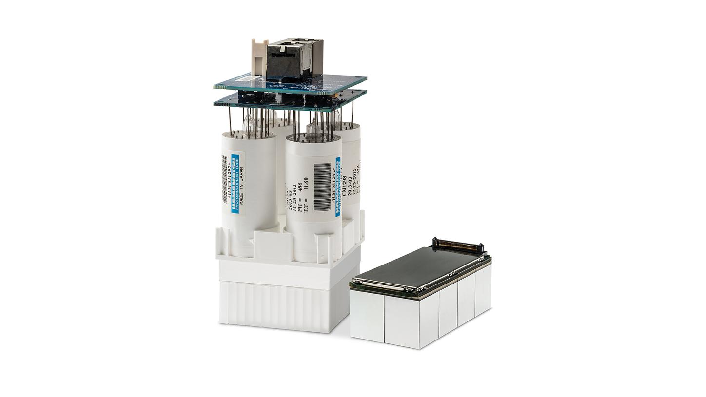
Optiso UDR detector
CT capability
iMAR
SAFIRE
Whole-body dynamic imaging
QualityGuard™
FlowMotion AI™
FAST PET Workflow AI
Multiparametric PET Suite AI
OncoFreeze™ / OncoFreeze AI
Exclusive bed design and wide bore
Zero differential deflection for accurate attenuation correction and TG-66 compliant along with a 78-cm bore to support bariatric imaging and easier positioning of radiation oncology devices
"…by improving the spatial resolution…you have less partial volume effect, so you get sharper images and more accurate quantification."

Ronald Boellaard, PhD
"We have now already, as compared to the older system, reduced the activity we inject… Now it's probably 30% faster with about 30% less dose which is something very acceptable."2

John Prior, PhD, MD
"Being more quantitative, our reproducibility can be that much better, and it may matter when we’re trying to do a repeat scan early on in a therapy and decide what to do."

Watch the full-length documentary video on how Biograph Vision became a reality.
Case 1
02Case 2
04Case 3
04Case 4
04Case 5
02Case 6
03Case 7
03Case 8
03


Visualization of vascular blood pool during whole-body dynamic PET/CT acquisition


Case 3
Visualization of 7 mm lymph node metastases on 68Ga-PSMA[a] evaluation of primary prostate cancer

Case 3
Visualization of 7 mm lymph node metastases on 68Ga-PSMA[a] evaluation of primary prostate cancer

Case 3
Visualization of 7 mm lymph node metastases on 68Ga-PSMA[a] evaluation of primary prostate cancer

Case 3
Visualization of 7 mm lymph node metastases on 68Ga-PSMA[a] evaluation of primary prostate cancer


Case 5
Small liver metastases in a patient with operated colorectal carcinoma detected with 18F FDG* PET/CT

Case 5
Small liver metastases in a patient with operated colorectal carcinoma detected with 18F FDG* PET/CT


Case 6
18F-Fluroestradiol[a] PET/CT in a patient with breast carcinoma


Case 7
Sharp delineation of physiological uptake in vascular walls, marrow, and intestine with 18F FDG* PET/CT

Case 7
Sharp delineation of physiological uptake in vascular walls, marrow, and intestine with 18F FDG* PET/CT

Case 7
Sharp delineation of physiological uptake in vascular walls, marrow, and intestine with 18F FDG* PET/CT


Visualization of vascular blood pool during whole-body dynamic PET/CT acquisition


Case 3
Visualization of 7 mm lymph node metastases on 68Ga-PSMA[a] evaluation of primary prostate cancer

Case 3
Visualization of 7 mm lymph node metastases on 68Ga-PSMA[a] evaluation of primary prostate cancer

Case 3
Visualization of 7 mm lymph node metastases on 68Ga-PSMA[a] evaluation of primary prostate cancer

Case 3
Visualization of 7 mm lymph node metastases on 68Ga-PSMA[a] evaluation of primary prostate cancer


Case 5
Small liver metastases in a patient with operated colorectal carcinoma detected with 18F FDG* PET/CT

Case 5
Small liver metastases in a patient with operated colorectal carcinoma detected with 18F FDG* PET/CT


Case 6
18F-Fluroestradiol[a] PET/CT in a patient with breast carcinoma


Case 7
Sharp delineation of physiological uptake in vascular walls, marrow, and intestine with 18F FDG* PET/CT

Case 7
Sharp delineation of physiological uptake in vascular walls, marrow, and intestine with 18F FDG* PET/CT

Case 7
Sharp delineation of physiological uptake in vascular walls, marrow, and intestine with 18F FDG* PET/CT


Transcend digital with the Optiso UDR detector
Optiso UDR’s proprietary 3.2-mm LSO crystals move silicon photomultiplier (SiPM) technology beyond digital to a new level of precision to help you detect small lesions, devise accurate treatment strategies\, and achieve optimal performance in a wide range of count rates.

See how Optiso Ultra Dynamic Range (UDR) PET detector advances PET/CT beyond digital.
Gantry | |
Bore diameter | 78 cm |
Tunnel length | 136 cm |
Table capacity | 227 kg (500 lb) |
CT | |
Generator power | 80 kW (100 kW optional) |
Rotation times | 0.33, 0.305, 0.285 s |
Tube voltages | 70, 80, 100, 120, 140 kV |
Iterative reconstruction | SAFIRE5 |
Metal artifact reduction | iMAR5 |
Slices | 64, 128 |
PET | |
Axial field of view | 26.3 cm |
Crystal size | 3.2 x 3.2 x 20 mm |
SiPM coverage of crystal array | 100% |
Effective sensitivity | 100 cps/kBq |
Effective NEC | 1870 kcps |
Time of flight performance | 214 ps |
Biograph Vision is not commercially available in all countries. Due to regulatory reasons, its future availability cannot be guaranteed. Please contact your local Siemens organization for further details.
Tyto stránky jsou určeny odborným pracovníkům ve zdravotnictví. Informace nejsou určeny pro laickou veřejnost.
Potvrzuji, že jsem odborníkem ve smyslu §2 a Zákona č. 40/1995 Sb., o regulaci reklamy, ve znění pozdějších předpisů, čili jsem osobou oprávněnou předepisovat léčivé přípravky nebo osobou oprávněnou léčivé přípravky vydávat.
Beru na vědomí, že informace obsažené dále na těchto stránkách nejsou určeny laické veřejnosti, nýbrž zdravotnickým odborníkům, a to se všemi riziky a důsledky z toho plynoucími pro laickou veřejnost.
