- Home
- Medical Imaging
- Molekulare Bildgebung
- Molecular Imaging Clinical Corner
- Clinical image galleries
- Biograph Vision PET/CT clinical image gallery
Biograph Vision PET/CT
Clinical gallery

Select a clinical case to review
Overview
01Case 2
Case 3
Case 4
Case 5
Case 6
Case 7
Case 8
Case 9
Case 10
Case 11
Data courtesy of University Medical Center Groningen, Groningen, The Netherlands.
Case 1: High-resolution reconstruction provides precise anatomical detail in normal 18F FDG brain study
- Small crystal elements in high image quality
- In brain imaging, this translates to precise contrast between white and gray matter
- Sulci, gyri, and mid-brain structures are well delineated
PET - Biograph Vision™ 600
Scan acquisition: 440 x 440 matrix, PSF+TOF 8i5s
Scan time: 15 minutes (1 bed position / 15 minutes per bed)
Injected dose: Fludeoxyglucose F 18 (18F FDG) Injection;
5.4 mCi (200 MBq)
CT
SAFIRE
Scan parameters: 80 kV/30 ref mAs
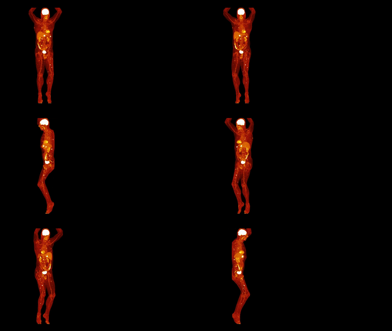
Case 2: FlowMotion-enabled whole-body
18F FDHT dynamic acquisition provides visualization of vascular blood pool
- Sharp delineation of vascular structures and biliary tree reflecting high system resolution
- Consistent image quality across all whole-body passes suggests potential for fast imaging
PET - Biograph Vision™ 600
Scan acquisition: 220 x 220 matrix, PSF+TOF 4i5s
Scan time: Dynamic acquisition - 6 whole-body passes;
2 minutes 4 seconds per pass
Injected dose: Fluoro-dihydrotestosterone (18F FDHT)[a];
7.5 mCi (200 MBq)
CT
SAFIRE
Scan parameters: 100 kV/13 ref mAs

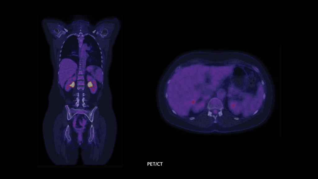
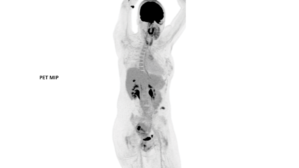
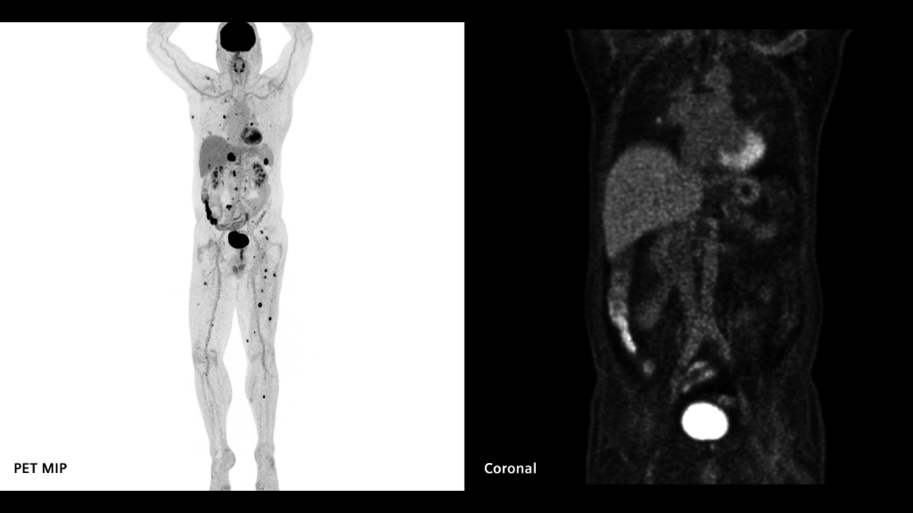
Case 3: 3.2 mm crystals and ultraHD•PET yield high image contrast in pelvic nodal and hepatic metastases in ovarian carcinoma
- Small crystals yield images of high resolution, allowing detection of small lesions
- ultraHD•PET results in heightened contrast of lesion to background
PET - Biograph Vision™ 600
Scan acquisition: 220 x 220 matrix, PSF+TOF 3i5s
Scan time: 21 minutes (7 bed positions / 3 minutes per bed)
Injected dose: Fludeoxyglucose F 18 (18F FDG) Injection;
7.5 mCi (280 MBq)
CT
SAFIRE
Scan parameters: 100 kV/16 ref mAs
Data courtesy of Centre Hospitalier Universitaire Vaudois, Lausanne, Switzerland.
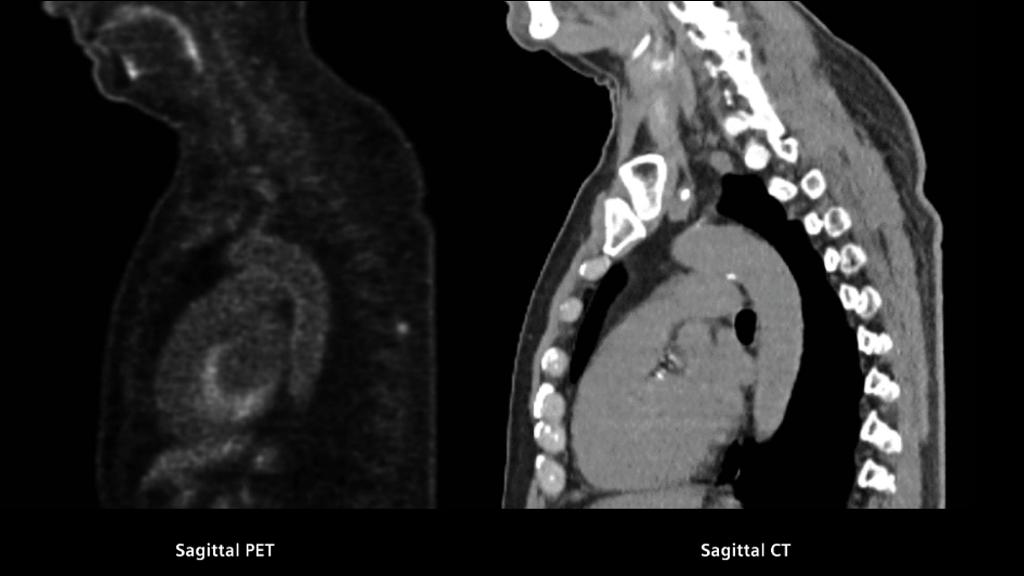
Case 4: SiPM coupled to 3.2 mm crystals sharply defines multiple metastases in myeloma
- Silicon photomultipliers (SiPM) and small crystal elements result in precise anatomical detail
- The walls of medium- and large- caliber vasculature are well-defined
- Detailed definition of mesentery from intra-abdominal organs and subcutaneous fat from underlying muscle
PET - Biograph Vision™ 600
Scan acquisition: 440 x 440 matrix, PSF+TOF 5i5s
Scan time: 27 minutes (9 bed positions / 3 minutes per bed)
Injected dose: Fludeoxyglucose F 18 (18F FDG) Injection;
7.4 mCi (274 MBq)
CT
SAFIRE
Scan parameters: 100 kV/125 ref mAs

Data courtesy of University Medical Center Groningen, Groningen, The Netherlands.
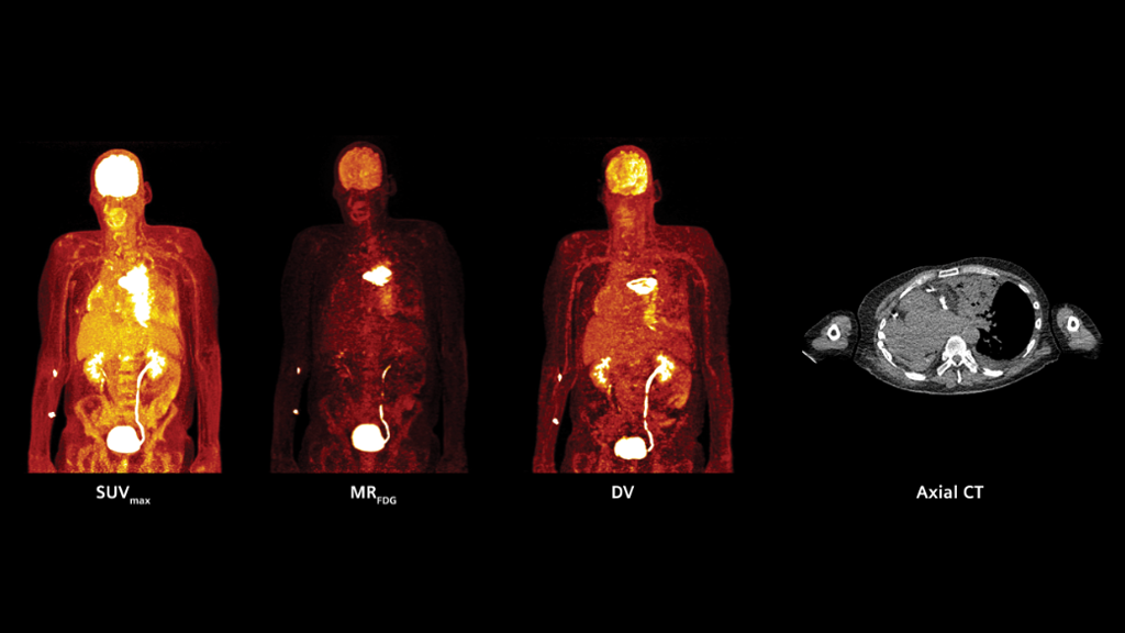
Case 5: FlowMotion Multiparametric PET AI helps physicians differentiate active tumor from induced-tissue effects
- The SUVmax image shows increased activity throughout the entire left lung mass
- The MRFDG and DV images demonstrate increased activity in the upper portion of the lesion, reflecting actively metabolizing tumor
- The lower portion, however, has only mild activity likely reflecting post-obstructive changes, which correlates to CT findings
- Metabolic rate of FDG is significantly different between tumor and induced-tissue effects
PET - Biograph Vision™ 600
Scan acquisition: 440 x 440 matrix, PSF+TOF 4i5s
Scan time: 60 minutes (dynamic acquisition - 19 whole-body passes)
Injected dose: Fludeoxyglucose F 18 (18F FDG) Injection;
6.32 mCi (234 MBq)
CT
SAFIRE
Scan parameters: 100 kV/11 ref mAs
Data courtesy of University Medical Center Groningen, Groningen, The Netherlands.
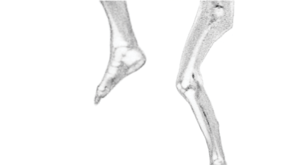
Case 6: SiPM along with 3.2 mm crystals enhances precise anatomical detail even from functional imaging
- 3.2 mm crystals along with silicon photomultipliers (SiPM) yield high-resolution images
- The result is precise anatomical detail:
- Small joints very well delineated
- Walls of medium and large caliber vessels well defined
Scan acquisition: 440 x 440 matrix, PSF+TOF 4i5s
Total scan time: 27 minutes (9 bed positions / 3 minutes per bed)
Injected dose: Fludeoxyglucose F 18 (18F FDG) Injection;
7.5 mCi (280 MBq)
CT
SAFIRE
Scan parameters: 120 kV/10 ref mAs
Data courtesy of University Medical Center Groningen, Groningen, The Netherlands./
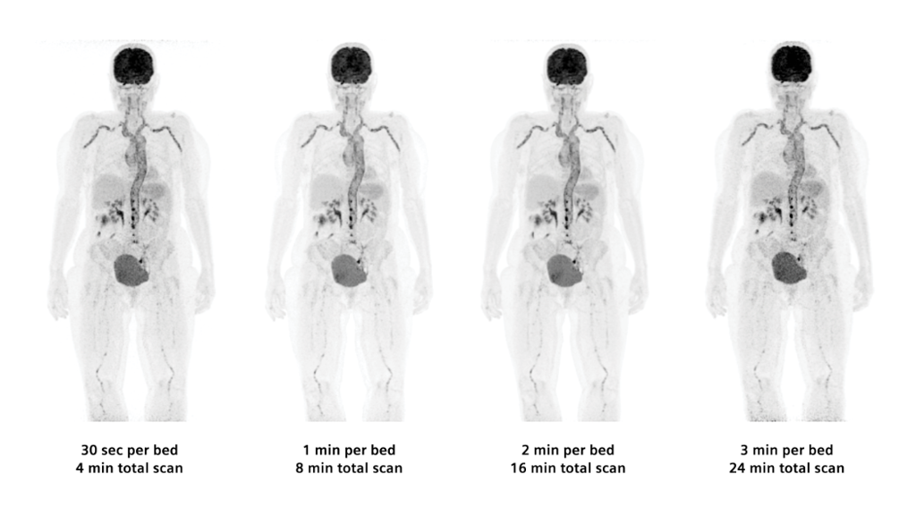
Case 7: Time-of-flight performance enables outstanding delineation of vasculature even at 30 seconds per bed
- There is high image resolution due to small crystal elements, strong signal due to optimal light capture by silicon photomultipliers (SiPM), and enhanced image contrast due to ultraHD•PET reconstruction
- The overall result is an image with precise anatomical detail even when acquired at low doses and fast scan times
PET - Biograph Vision™ 600
Scan acquisition: 440 x 440 matrix, PSF+TOF 4i5s
Scan time: 8 bed positions / 30 seconds, 1, 2, and 3 minutes per bed
Injected dose: Fludeoxyglucose F 18 (18F FDG) Injection;
4.1 mCi (150 MBq)
SAFIRE
Scan parameters: 120 kV/9 ref mAs
Data courtesy of Centre Hospitalier Universitaire Vaudois, Lausanne, Switzerland.
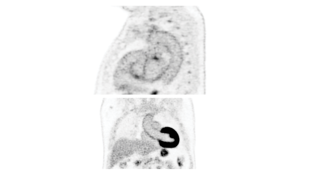
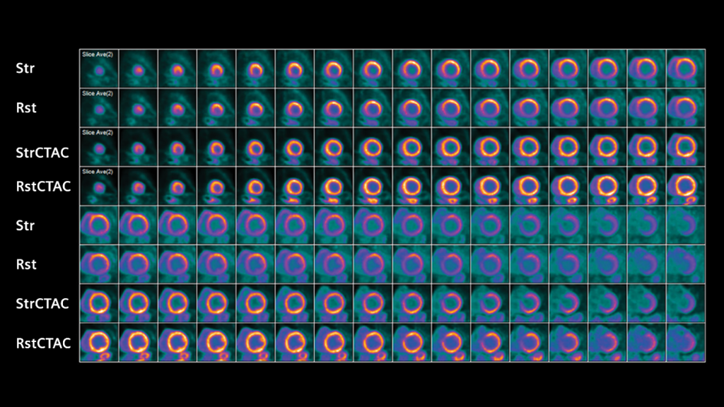
Case 8: Time of flight and high resolution enable low-dose (18.9 mCi) 82Rb study with high image quality
- 3.2 mm LSO crystals mean better definition of myocardial wall/intraventricular cavity
- Precise images with less than half of the conventional 82Rb (40 mCi/1480 MBq) dose
- Accurate myocardial blood flow (MBF) quantification as detection saturation is not an issue
PET - Biograph Vision™ 600
Scan acquisition: 220 x 220 matrix, PSF+TOF 4i5s
Scan time: 6 minutes (1 bed position / 6 minutes per bed)
Injected dose: Rubidium 82 (82Rb) 18.9 mCi (700 MBq)
SAFIRE
Scan parameters: 100 kV/9 ref mAs

Data courtesy of Centre Hospitalier Régional et Universitaire de Brest, Brest, France.
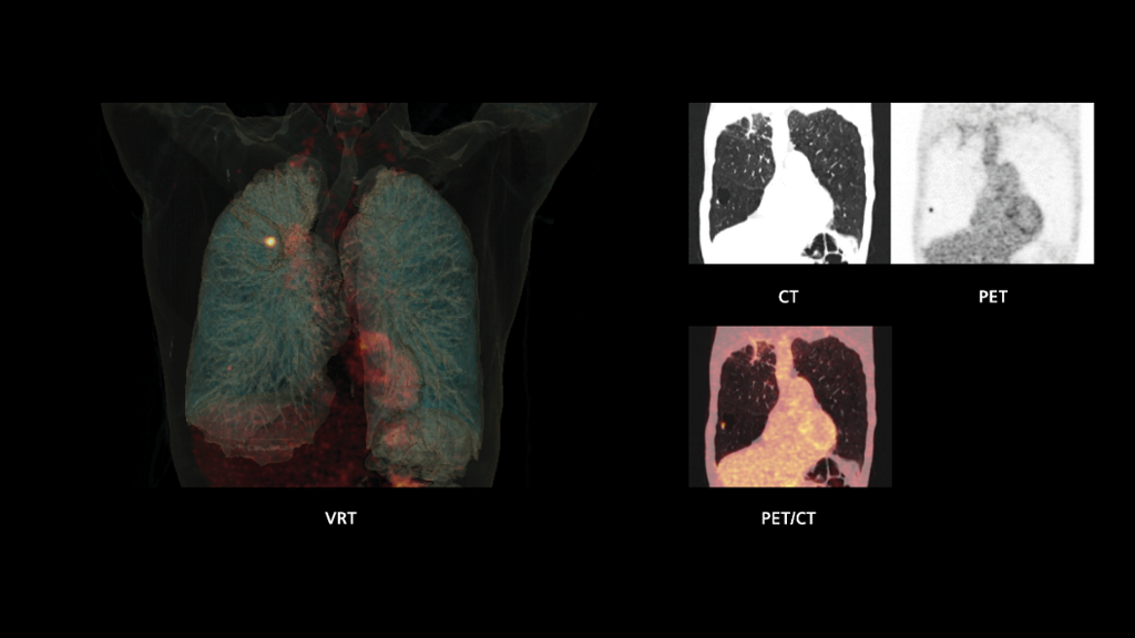
Case 9: Small crystal size allows precise visualization of a 6 mm lung nodule with 2.76 mCi (102 MBq) acquired in 6 minutes
- Sharp delineation of small pulmonary nodule near the right lung base acquired with low dose and short acquisition time
- High resolution and contrast of PET images obtained because of small crystals and high time-of-flight (ToF) performance enable high-sensitivity acquisitions providing uncompromised image quality even at low dose
PET - Biograph Vision™ 600
Scan acquisition: 440 x 440 matrix, PSF+TOF 3i5s
Scan time: 6 minutes
Injected dose: Fludeoxyglucose F 18 (18F FDG) Injection;
2.76 mCi (102 MBq)
CT
SAFIRE
Scan parameters: 100 kV/18 ref mAs
Data courtesy of University Medical Center Groningen, Groningen, The Netherlands.
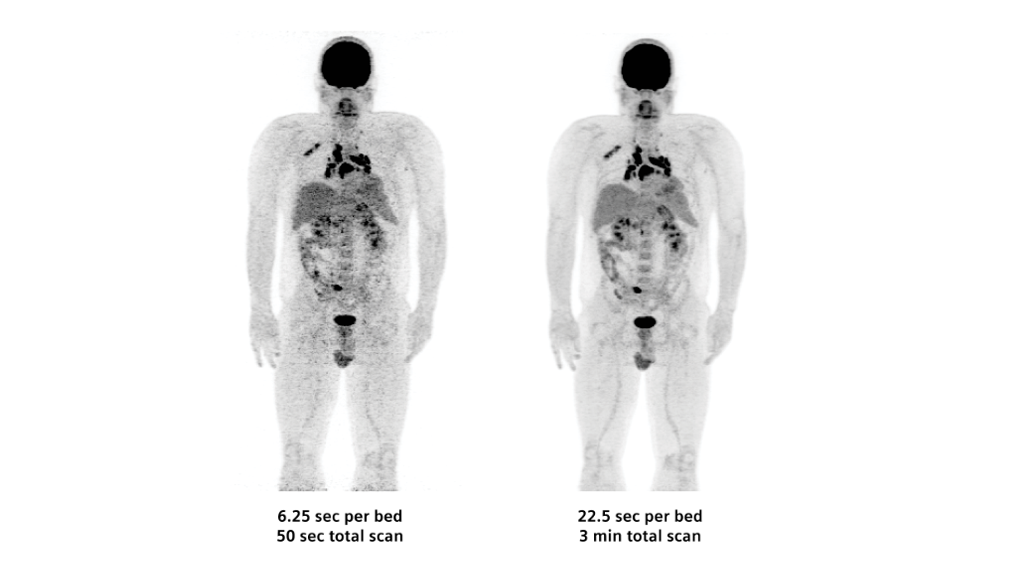
Case 10: 3.2 mm crystals along with ultraHD•PET allows low-dose and fast (50 seconds) total whole-body scan
- 50-second, whole-body acquisition reveals extent of disease
- Images show high resolution and contrast-to-noise ratio due to 3.2 mm crystal elements and ultraHD•PET reconstruction, which helps enable fast scan times and low doses
Scan acquisition: 220 x 220 matrix, PSF+TOF 1i5s, Gaussian filter 2
Scan time: Whole-body 8 bed positions (6.25 and 22.5 seconds per bed)
Injected dose: Fludeoxyglucose F 18 (18F FDG) Injection;
5.9 mCi (220 MBq)
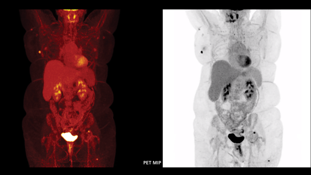
Data courtesy of Indiana University Methodist Hospital, Indiana, USA.
Case 11: Time-of-flight performance enables precise image quality in an 107 kg (236 lb) obese patient
- Small lesions easily characterized in obese patients as a result of ultraHD•PET
- FlowMotion™ continuous-bed-motion technology allows image acquisition flexibility with high image resolution in a designated region
PET - Biograph Vision™ 600
Scan acquisition: 440 x 440 matrix, PSF+TOF 3i5s, All-pass filter
Scan time: FlowMotion™ continuous-bed-motion;
Head to pelvis at 1.0 mm/s
Injected dose: Fludeoxyglucose F 18 (18F FDG) Injection;
17 mCi (640 MBq)
CT
Scan parameters: 120 kV/110 ref mAs
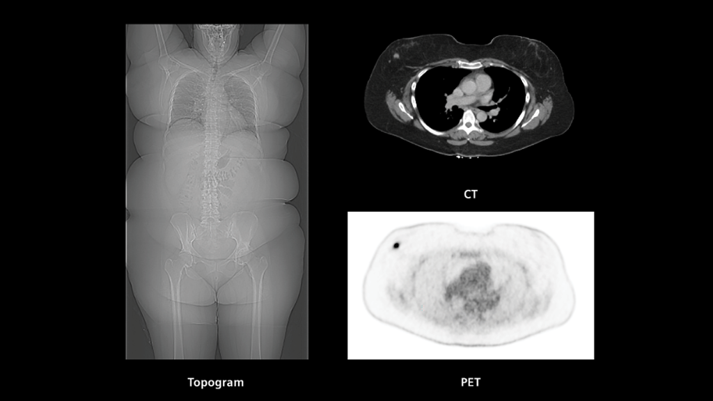
Data courtesy of Naestved Hospital, Naestved, Denmark.
Case 12: iMAR helps reduce metal artifacts from hip prosthesis in CT to enable clear visualization of uptake in pelvic wall
- CT with iterative algorithm for metal artifact reduction (iMAR) reconstruction reduces metal artifacts from bilateral hip prosthesis in a patient with pathology around the right hip prosthesis
- Sharp and clear delineation of uptake in the pelvic wall margin adjacent to right prosthetic hip joint is made possible due to artifact-free CT with improved soft-tissue delineation
PET - Biograph Vision™ 450
Scan acquisition: 440 x 440 matrix, PSF+TOF 4i5s, All-pass filter
Scan time: 16.9 minutes; FlowMotion™ continuous-bed-motion;
1 zone 1.0 mm/s
Injected dose: Fludeoxyglucose F 18 (18F FDG) Injection;
8.7 mCi (322 MBq)
CT
SAFIRE
Scan parameters: 100 kV/280 ref mAs
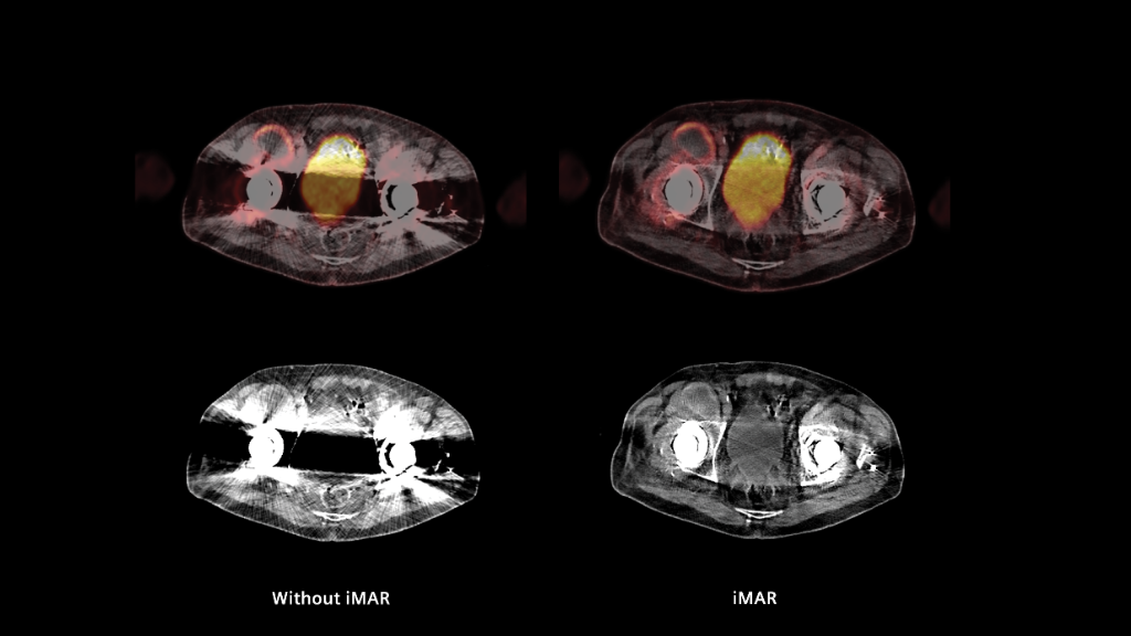
Data courtesy of Naestved Hospital, Naestved, Denmark.
Case 13: ToF performance and high-resolution PET provide sharp detail of physiological colonic wall uptake
- Silicon photomultipliers (SiPM)-related time-of-flight (ToF) performance (214 picoseconds) and high-resolution PET due to 3.2 mm crystal size enable sharp visualization of physiological uptake in the entire colonic wall, as well as renal calyces, pelvis, and the entire ureter
- High image quality with low background reflecting high count statistics related to ToF performance
PET - Biograph Vision™ 450
Scan acquisition: 440 x 440 matrix, PSF+TOF 4i5s, All-pass filter
Scan time: 16.8 minutes; FlowMotion™ continuous-bed-motion;
1 zone 1.0 mm/s
Injected dose: Fludeoxyglucose F 18 (18F FDG) Injection;
10 mCi (375 MBq)
CT
SAFIRE
Scan parameters: 120 kV/38 ref mAs

Data and images courtesy of Inselspital, Universitätsspital Bern, Switzerland.
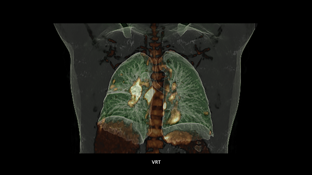
Case 14: Single-bed, whole-body acquisition in lung cancer
- Lung cancer (post-COVID-19)
- 140-minute uptake
- 173 cm (5'6")
- 95 kg (209 lb)
PET - Biograph Vision Quadra™
Scan acquisition: 440 x 440 matrix, PSF+TOF 4i5s, Gaussian filter 2
Scan time: 10 minutes (1 bed position / 10 minutes per bed)
Injected dose: Fludeoxyglucose F 18 (18F FDG) Injection;
10.1 mCi (373 MBq)
CT
Scan parameters: 100 kV/20 ref mAs
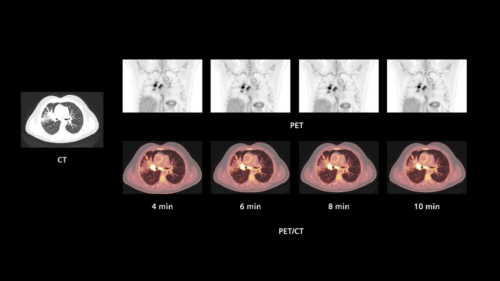
Data courtesy of University Medical Center Groningen, Groningen, The Netherlands.
Case 1: High-resolution reconstruction provides precise anatomical detail in normal 18F FDG brain study
- Small crystal elements in high image quality
- In brain imaging, this translates to precise contrast between white and gray matter
- Sulci, gyri, and mid-brain structures are well delineated
PET - Biograph Vision™ 600
Scan acquisition: 440 x 440 matrix, PSF+TOF 8i5s
Scan time: 15 minutes (1 bed position / 15 minutes per bed)
Injected dose: Fludeoxyglucose F 18 (18F FDG) Injection;
5.4 mCi (200 MBq)
CT
SAFIRE
Scan parameters: 80 kV/30 ref mAs
Case 2: FlowMotion-enabled whole-body
18F FDHT dynamic acquisition provides visualization of vascular blood pool
- Sharp delineation of vascular structures and biliary tree reflecting high system resolution
- Consistent image quality across all whole-body passes suggests potential for fast imaging
PET - Biograph Vision™ 600
Scan acquisition: 220 x 220 matrix, PSF+TOF 4i5s
Scan time: Dynamic acquisition - 6 whole-body passes;
2 minutes 4 seconds per pass
Injected dose: Fluoro-dihydrotestosterone (18F FDHT)[a];
7.5 mCi (200 MBq)
CT
SAFIRE
Scan parameters: 100 kV/13 ref mAs

Case 3: 3.2 mm crystals and ultraHD•PET yield high image contrast in pelvic nodal and hepatic metastases in ovarian carcinoma
- Small crystals yield images of high resolution, allowing detection of small lesions
- ultraHD•PET results in heightened contrast of lesion to background
PET - Biograph Vision™ 600
Scan acquisition: 220 x 220 matrix, PSF+TOF 3i5s
Scan time: 21 minutes (7 bed positions / 3 minutes per bed)
Injected dose: Fludeoxyglucose F 18 (18F FDG) Injection;
7.5 mCi (280 MBq)
CT
SAFIRE
Scan parameters: 100 kV/16 ref mAs

Data courtesy of Centre Hospitalier Universitaire Vaudois, Lausanne, Switzerland.
Case 4: SiPM coupled to 3.2 mm crystals sharply defines multiple metastases in myeloma
- Silicon photomultipliers (SiPM) and small crystal elements result in precise anatomical detail
- The walls of medium- and large- caliber vasculature are well-defined
- Detailed definition of mesentery from intra-abdominal organs and subcutaneous fat from underlying muscle
PET - Biograph Vision™ 600
Scan acquisition: 440 x 440 matrix, PSF+TOF 5i5s
Scan time: 27 minutes (9 bed positions / 3 minutes per bed)
Injected dose: Fludeoxyglucose F 18 (18F FDG) Injection;
7.4 mCi (274 MBq)
CT
SAFIRE
Scan parameters: 100 kV/125 ref mAs

Data courtesy of University Medical Center Groningen, Groningen, The Netherlands.
Case 5: FlowMotion Multiparametric PET AI helps physicians differentiate active tumor from induced-tissue effects
- The SUVmax image shows increased activity throughout the entire left lung mass
- The MRFDG and DV images demonstrate increased activity in the upper portion of the lesion, reflecting actively metabolizing tumor
- The lower portion, however, has only mild activity likely reflecting post-obstructive changes, which correlates to CT findings
- Metabolic rate of FDG is significantly different between tumor and induced-tissue effects
PET - Biograph Vision™ 600
Scan acquisition: 440 x 440 matrix, PSF+TOF 4i5s
Scan time: 60 minutes (dynamic acquisition - 19 whole-body passes)
Injected dose: Fludeoxyglucose F 18 (18F FDG) Injection;
6.32 mCi (234 MBq)
CT
SAFIRE
Scan parameters: 100 kV/11 ref mAs

Data courtesy of University Medical Center Groningen, Groningen, The Netherlands.
Case 5: FlowMotion Multiparametric PET AI helps physicians differentiate active tumor from induced-tissue effects
- The SUVmax image shows increased activity throughout the entire left lung mass
- The MRFDG and DV images demonstrate increased activity in the upper portion of the lesion, reflecting actively metabolizing tumor
- The lower portion, however, has only mild activity likely reflecting post-obstructive changes, which correlates to CT findings
- Metabolic rate of FDG is significantly different between tumor and induced-tissue effects
PET - Biograph Vision™ 600
Scan acquisition: 440 x 440 matrix, PSF+TOF 4i5s
Scan time: 60 minutes (dynamic acquisition - 19 whole-body passes)
Injected dose: Fludeoxyglucose F 18 (18F FDG) Injection;
6.32 mCi (234 MBq)
CT
SAFIRE
Scan parameters: 100 kV/11 ref mAs

Data courtesy of University Medical Center Groningen, Groningen, The Netherlands.
Case 6: SiPM along with 3.2 mm crystals enhances precise anatomical detail even from functional imaging
- 3.2 mm crystals along with silicon photomultipliers (SiPM) yield high-resolution images
- The result is precise anatomical detail:
- Small joints very well delineated
- Walls of medium and large caliber vessels well defined
Scan acquisition: 440 x 440 matrix, PSF+TOF 4i5s
Total scan time: 27 minutes (9 bed positions / 3 minutes per bed)
Injected dose: Fludeoxyglucose F 18 (18F FDG) Injection;
7.5 mCi (280 MBq)
CT
SAFIRE
Scan parameters: 120 kV/10 ref mAs

Data courtesy of University Medical Center Groningen, Groningen, The Netherlands./
Case 7: Time-of-flight performance enables outstanding delineation of vasculature even at 30 seconds per bed
- There is high image resolution due to small crystal elements, strong signal due to optimal light capture by silicon photomultipliers (SiPM), and enhanced image contrast due to ultraHD•PET reconstruction
- The overall result is an image with precise anatomical detail even when acquired at low doses and fast scan times
PET - Biograph Vision™ 600
Scan acquisition: 440 x 440 matrix, PSF+TOF 4i5s
Scan time: 8 bed positions / 30 seconds, 1, 2, and 3 minutes per bed
Injected dose: Fludeoxyglucose F 18 (18F FDG) Injection;
4.1 mCi (150 MBq)
SAFIRE
Scan parameters: 120 kV/9 ref mAs

Data courtesy of Centre Hospitalier Universitaire Vaudois, Lausanne, Switzerland.
Case 8: Time of flight and high resolution enable low-dose (18.9 mCi) 82Rb study with high image quality
- 3.2 mm LSO crystals mean better definition of myocardial wall/intraventricular cavity
- Precise images with less than half of the conventional 82Rb (40 mCi/1480 MBq) dose
- Accurate myocardial blood flow (MBF) quantification as detection saturation is not an issue
PET - Biograph Vision™ 600
Scan acquisition: 220 x 220 matrix, PSF+TOF 4i5s
Scan time: 6 minutes (1 bed position / 6 minutes per bed)
Injected dose: Rubidium 82 (82Rb) 18.9 mCi (700 MBq)
SAFIRE
Scan parameters: 100 kV/9 ref mAs

Data courtesy of Centre Hospitalier Universitaire Vaudois, Lausanne, Switzerland.
Case 8: Time of flight and high resolution enable low-dose (18.9 mCi) 82Rb study with high image quality
- 3.2 mm LSO crystals mean better definition of myocardial wall/intraventricular cavity
- Precise images with less than half of the conventional 82Rb (40 mCi/1480 MBq) dose
- Accurate myocardial blood flow (MBF) quantification as detection saturation is not an issue
PET - Biograph Vision™ 600
Scan acquisition: 220 x 220 matrix, PSF+TOF 4i5s
Scan time: 6 minutes (1 bed position / 6 minutes per bed)
Injected dose: Rubidium 82 (82Rb) 18.9 mCi (700 MBq)
SAFIRE
Scan parameters: 100 kV/9 ref mAs

Data courtesy of Centre Hospitalier Universitaire Vaudois, Lausanne, Switzerland.
Case 8: Time of flight and high resolution enable low-dose (18.9 mCi) 82Rb study with high image quality
- 3.2 mm LSO crystals mean better definition of myocardial wall/intraventricular cavity
- Precise images with less than half of the conventional 82Rb (40 mCi/1480 MBq) dose
- Accurate myocardial blood flow (MBF) quantification as detection saturation is not an issue
PET - Biograph Vision™ 600
Scan acquisition: 220 x 220 matrix, PSF+TOF 4i5s
Scan time: 6 minutes (1 bed position / 6 minutes per bed)
Injected dose: Rubidium 82 (82Rb) 18.9 mCi (700 MBq)
SAFIRE
Scan parameters: 100 kV/9 ref mAs

Data courtesy of Centre Hospitalier Régional et Universitaire de Brest, Brest, France.
Case 9: Small crystal size allows precise visualization of a 6 mm lung nodule with 2.76 mCi (102 MBq) acquired in 6 minutes
- Sharp delineation of small pulmonary nodule near the right lung base acquired with low dose and short acquisition time
- High resolution and contrast of PET images obtained because of small crystals and high time-of-flight (ToF) performance enable high-sensitivity acquisitions providing uncompromised image quality even at low dose
PET - Biograph Vision™ 600
Scan acquisition: 440 x 440 matrix, PSF+TOF 3i5s
Scan time: 6 minutes
Injected dose: Fludeoxyglucose F 18 (18F FDG) Injection;
2.76 mCi (102 MBq)
CT
SAFIRE
Scan parameters: 100 kV/18 ref mAs
Data courtesy of Centre Hospitalier Régional et Universitaire de Brest, Brest, France.
Case 9: Small crystal size allows precise visualization of a 6 mm lung nodule with 2.76 mCi (102 MBq) acquired in 6 minutes
- Sharp delineation of small pulmonary nodule near the right lung base acquired with low dose and short acquisition time
- High resolution and contrast of PET images obtained because of small crystals and high time-of-flight (ToF) performance enable high-sensitivity acquisitions providing uncompromised image quality even at low dose
PET - Biograph Vision™ 600
Scan acquisition: 440 x 440 matrix, PSF+TOF 3i5s
Scan time: 6 minutes
Injected dose: Fludeoxyglucose F 18 (18F FDG) Injection;
2.76 mCi (102 MBq)
CT
SAFIRE
Scan parameters: 100 kV/18 ref mAs

Data courtesy of Centre Hospitalier Régional et Universitaire de Brest, Brest, France.
Case 9: Small crystal size allows precise visualization of a 6 mm lung nodule with 2.76 mCi (102 MBq) acquired in 6 minutes
- Sharp delineation of small pulmonary nodule near the right lung base acquired with low dose and short acquisition time
- High resolution and contrast of PET images obtained because of small crystals and high time-of-flight (ToF) performance enable high-sensitivity acquisitions providing uncompromised image quality even at low dose
PET - Biograph Vision™ 600
Scan acquisition: 440 x 440 matrix, PSF+TOF 3i5s
Scan time: 6 minutes
Injected dose: Fludeoxyglucose F 18 (18F FDG) Injection;
2.76 mCi (102 MBq)
CT
SAFIRE
Scan parameters: 100 kV/18 ref mAs

Data courtesy of University Medical Center Groningen, Groningen, The Netherlands.
Case 10: 3.2 mm crystals along with ultraHD•PET allows low-dose and fast (50 seconds) total whole-body scan
- 50-second, whole-body acquisition reveals extent of disease
- Images show high resolution and contrast-to-noise ratio due to 3.2 mm crystal elements and ultraHD•PET reconstruction, which helps enable fast scan times and low doses
Scan acquisition: 220 x 220 matrix, PSF+TOF 1i5s, Gaussian filter 2
Scan time: Whole-body 8 bed positions (6.25 and 22.5 seconds per bed)
Injected dose: Fludeoxyglucose F 18 (18F FDG) Injection;
5.9 mCi (220 MBq)

Data courtesy of Indiana University Methodist Hospital, Indiana, USA.
Case 11: Time-of-flight performance enables precise image quality in an 107 kg (236 lb) obese patient
- Small lesions easily characterized in obese patients as a result of ultraHD•PET
- FlowMotion™ continuous-bed-motion technology allows image acquisition flexibility with high image resolution in a designated region
PET - Biograph Vision™ 600
Scan acquisition: 440 x 440 matrix, PSF+TOF 3i5s, All-pass filter
Scan time: FlowMotion™ continuous-bed-motion;
Head to pelvis at 1.0 mm/s
Injected dose: Fludeoxyglucose F 18 (18F FDG) Injection;
17 mCi (640 MBq)
CT
Scan parameters: 120 kV/110 ref mAs
Data courtesy of Indiana University Methodist Hospital, Indiana, USA.
Case 11: Time-of-flight performance enables precise image quality in an 107 kg (236 lb) obese patient
- Small lesions easily characterized in obese patients as a result of ultraHD•PET
- FlowMotion™ continuous-bed-motion technology allows image acquisition flexibility with high image resolution in a designated region
PET - Biograph Vision™ 600
Scan acquisition: 440 x 440 matrix, PSF+TOF 3i5s, All-pass filter
Scan time: FlowMotion™ continuous-bed-motion;
Head to pelvis at 1.0 mm/s
Injected dose: Fludeoxyglucose F 18 (18F FDG) Injection;
17 mCi (640 MBq)
CT
Scan parameters: 120 kV/110 ref mAs

Data courtesy of Indiana University Methodist Hospital, Indiana, USA.
Case 11: Time-of-flight performance enables precise image quality in an 107 kg (236 lb) obese patient
- Small lesions easily characterized in obese patients as a result of ultraHD•PET
- FlowMotion™ continuous-bed-motion technology allows image acquisition flexibility with high image resolution in a designated region
PET - Biograph Vision™ 600
Scan acquisition: 440 x 440 matrix, PSF+TOF 3i5s, All-pass filter
Scan time: FlowMotion™ continuous-bed-motion;
Head to pelvis at 1.0 mm/s
Injected dose: Fludeoxyglucose F 18 (18F FDG) Injection;
17 mCi (640 MBq)
CT
Scan parameters: 120 kV/110 ref mAs

Data courtesy of Indiana University Methodist Hospital, Indiana, USA.
Case 11: Time-of-flight performance enables precise image quality in an 107 kg (236 lb) obese patient
- Small lesions easily characterized in obese patients as a result of ultraHD•PET
- FlowMotion™ continuous-bed-motion technology allows image acquisition flexibility with high image resolution in a designated region
PET - Biograph Vision™ 600
Scan acquisition: 440 x 440 matrix, PSF+TOF 3i5s, All-pass filter
Scan time: FlowMotion™ continuous-bed-motion;
Head to pelvis at 1.0 mm/s
Injected dose: Fludeoxyglucose F 18 (18F FDG) Injection;
17 mCi (640 MBq)
CT
Scan parameters: 120 kV/110 ref mAs
Data courtesy of Naestved Hospital, Naestved, Denmark.
Case 12: iMAR helps reduce metal artifacts from hip prosthesis in CT to enable clear visualization of uptake in pelvic wall
- CT with iterative algorithm for metal artifact reduction (iMAR) reconstruction reduces metal artifacts from bilateral hip prosthesis in a patient with pathology around the right hip prosthesis
- Sharp and clear delineation of uptake in the pelvic wall margin adjacent to right prosthetic hip joint is made possible due to artifact-free CT with improved soft-tissue delineation
PET - Biograph Vision™ 450
Scan acquisition: 440 x 440 matrix, PSF+TOF 4i5s, All-pass filter
Scan time: 16.9 minutes; FlowMotion™ continuous-bed-motion;
1 zone 1.0 mm/s
Injected dose: Fludeoxyglucose F 18 (18F FDG) Injection;
8.7 mCi (322 MBq)
CT
SAFIRE
Scan parameters: 100 kV/280 ref mAs

Data courtesy of Naestved Hospital, Naestved, Denmark.
Case 12: iMAR helps reduce metal artifacts from hip prosthesis in CT to enable clear visualization of uptake in pelvic wall
- CT with iterative algorithm for metal artifact reduction (iMAR) reconstruction reduces metal artifacts from bilateral hip prosthesis in a patient with pathology around the right hip prosthesis
- Sharp and clear delineation of uptake in the pelvic wall margin adjacent to right prosthetic hip joint is made possible due to artifact-free CT with improved soft-tissue delineation
PET - Biograph Vision™ 450
Scan acquisition: 440 x 440 matrix, PSF+TOF 4i5s, All-pass filter
Scan time: 16.9 minutes; FlowMotion™ continuous-bed-motion;
1 zone 1.0 mm/s
Injected dose: Fludeoxyglucose F 18 (18F FDG) Injection;
8.7 mCi (322 MBq)
CT
SAFIRE
Scan parameters: 100 kV/280 ref mAs
Data courtesy of Naestved Hospital, Naestved, Denmark.
Case 13: ToF performance and high-resolution PET provide sharp detail of physiological colonic wall uptake
- CT with iterative algorithm for metal artifact reduction (iMAR) reconstruction reduces metal artifacts from bilateral hip prosthesis in a patient with pathology around the right hip prosthesis
- Sharp and clear delineation of uptake in the pelvic wall margin adjacent to right prosthetic hip joint is made possible due to artifact-free CT with improved soft-tissue delineation
PET - Biograph Vision™ 450
Scan acquisition: 440 x 440 matrix, PSF+TOF 4i5s, All-pass filter
Scan time: 16.9 minutes; FlowMotion™ continuous-bed-motion;
1 zone 1.0 mm/s
Injected dose: Fludeoxyglucose F 18 (18F FDG) Injection;
8.7 mCi (322 MBq)
CT
SAFIRE
Scan parameters: 100 kV/280 ref mAs
Data courtesy of Naestved Hospital, Naestved, Denmark.
Case 13: ToF performance and high-resolution PET provide sharp detail of physiological colonic wall uptake
- Silicon photomultipliers (SiPM)-related time-of-flight (ToF) performance (214 picoseconds) and high-resolution PET due to 3.2 mm crystal size enable sharp visualization of physiological uptake in the entire colonic wall, as well as renal calyces, pelvis, and the entire ureter
- High image quality with low background reflecting high count statistics related to ToF performance
PET - Biograph Vision™ 450
Scan acquisition: 440 x 440 matrix, PSF+TOF 4i5s, All-pass filter
Scan time: 16.8 minutes; FlowMotion™ continuous-bed-motion;
1 zone 1.0 mm/s
Injected dose: Fludeoxyglucose F 18 (18F FDG) Injection;
10 mCi (375 MBq)
CT
SAFIRE
Scan parameters: 120 kV/38 ref mAs

Data and images courtesy of Inselspital, Universitätsspital Bern, Switzerland.
Case 14: Single-bed, whole-body acquisition in lung cancer
- Lung cancer (post-COVID-19)
- 140-minute uptake
- 173 cm (5'6")
- 95 kg (209 lb)
PET - Biograph Vision Quadra™
Scan acquisition: 440 x 440 matrix, PSF+TOF 4i5s, Gaussian filter 2
Scan time: 10 minutes (1 bed position / 10 minutes per bed)
Injected dose: Fludeoxyglucose F 18 (18F FDG) Injection;
10.1 mCi (373 MBq)
CT
Scan parameters: 100 kV/20 ref mAs

Data and images courtesy of Inselspital, Universitätsspital Bern, Switzerland.
Case 14: Single-bed, whole-body acquisition in lung cancer
- Lung cancer (post-COVID-19)
- 140-minute uptake
- 173 cm (5'6")
- 95 kg (209 lb)
PET - Biograph Vision Quadra™
Scan acquisition: 440 x 440 matrix, PSF+TOF 4i5s, Gaussian filter 2
Scan time: 10 minutes (1 bed position / 10 minutes per bed)
Injected dose: Fludeoxyglucose F 18 (18F FDG) Injection;
10.1 mCi (373 MBq)
CT
Scan parameters: 100 kV/20 ref mAs

Data and images courtesy of Inselspital, Universitätsspital Bern, Switzerland.
Case 14: Single-bed, whole-body acquisition in lung cancer
- Lung cancer (post-COVID-19)
- 140-minute uptake
- 173 cm (5'6")
- 95 kg (209 lb)
PET - Biograph Vision Quadra™
Scan acquisition: 440 x 440 matrix, PSF+TOF 4i5s, Gaussian filter 2
Scan time: 10 minutes (1 bed position / 10 minutes per bed)
Injected dose: Fludeoxyglucose F 18 (18F FDG) Injection;
10.1 mCi (373 MBq)
CT
Scan parameters: 100 kV/20 ref mAs

Data and images courtesy of Inselspital, Universitätsspital Bern, Switzerland.
Case 14: Single-bed, whole-body acquisition in lung cancer
- Lung cancer (post-COVID-19)
- 140-minute uptake
- 173 cm (5'6")
- 95 kg (209 lb)
PET - Biograph Vision Quadra™
Scan acquisition: 440 x 440 matrix, PSF+TOF 4i5s, Gaussian filter 2
Scan time: 10 minutes (1 bed position / 10 minutes per bed)
Injected dose: Fludeoxyglucose F 18 (18F FDG) Injection;
10.1 mCi (373 MBq)
CT
Scan parameters: 100 kV/20 ref mAs

Data and images courtesy of Inselspital, Universitätsspital Bern, Switzerland.
Case 14: Single-bed, whole-body acquisition in lung cancer
- Lung cancer (post-COVID-19)
- 140-minute uptake
- 173 cm (5'6")
- 95 kg (209 lb)
PET - Biograph Vision Quadra™
Scan acquisition: 440 x 440 matrix, PSF+TOF 4i5s, Gaussian filter 2
Scan time: 10 minutes (1 bed position / 10 minutes per bed)
Injected dose: Fludeoxyglucose F 18 (18F FDG) Injection;
10.1 mCi (373 MBq)
CT
Scan parameters: 100 kV/20 ref mAs


Did this information help you?
Thank you.
Biograph Vision and Biograph Vision Quadra are not commercially available in all countries. Future availability cannot be guaranteed. Please contact your local Siemens Healthineers organization for further details.
[a] The tracer used in this case is a research pharmaceutical. It is neither recognized to be safe and effective by the FDA nor commercially available in the United States or in other countries worldwide. Its future availability cannot be guaranteed. Please contact your local Siemens Healthineers organization for further details.