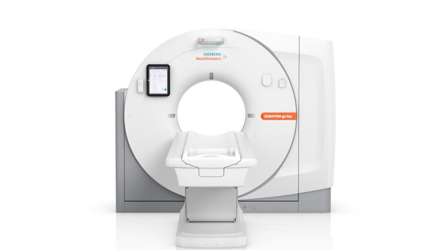
Patient pathways for guided CT imagingSOMATOM go.Top with myExam Companion
Patient pathways
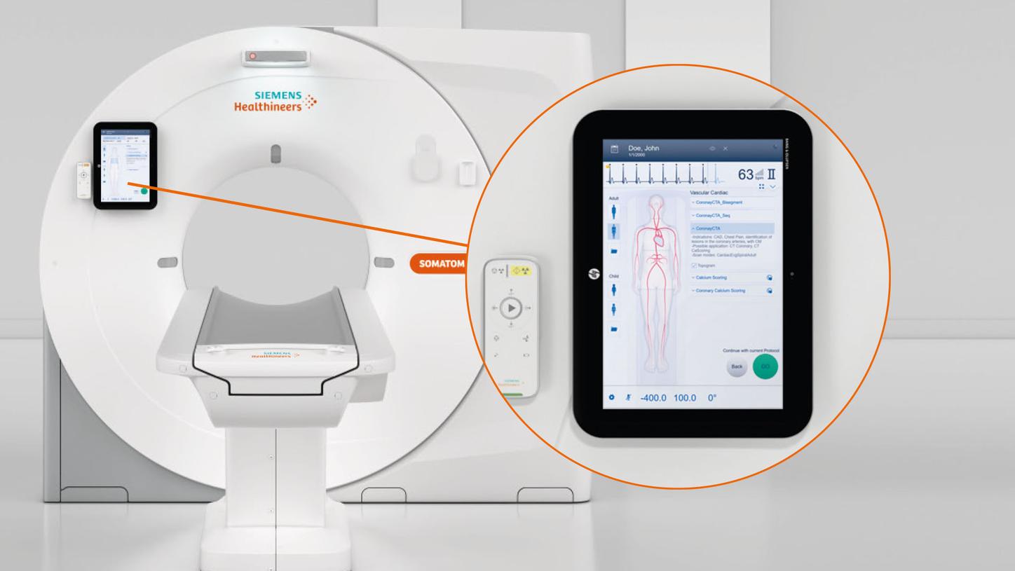
Transform care delivery with your new patient pathways
In CT imaging, each scan protocol contains many parameters. To attain optimal exam results, a subset of parameters needs to be manually adjusted for each patient and clinical indication. This can not only be time-consuming, but also result in inconsistent images because every staff member has a different level of experience and expertise – and their own work style.
Offering pre-defined patient pathways, myExam Companion easily navigates you through any clinical indication. Tap the full potential of the SOMATOM go.Top CT scanner and get consistent imaging results – independent of patient, case, and user.

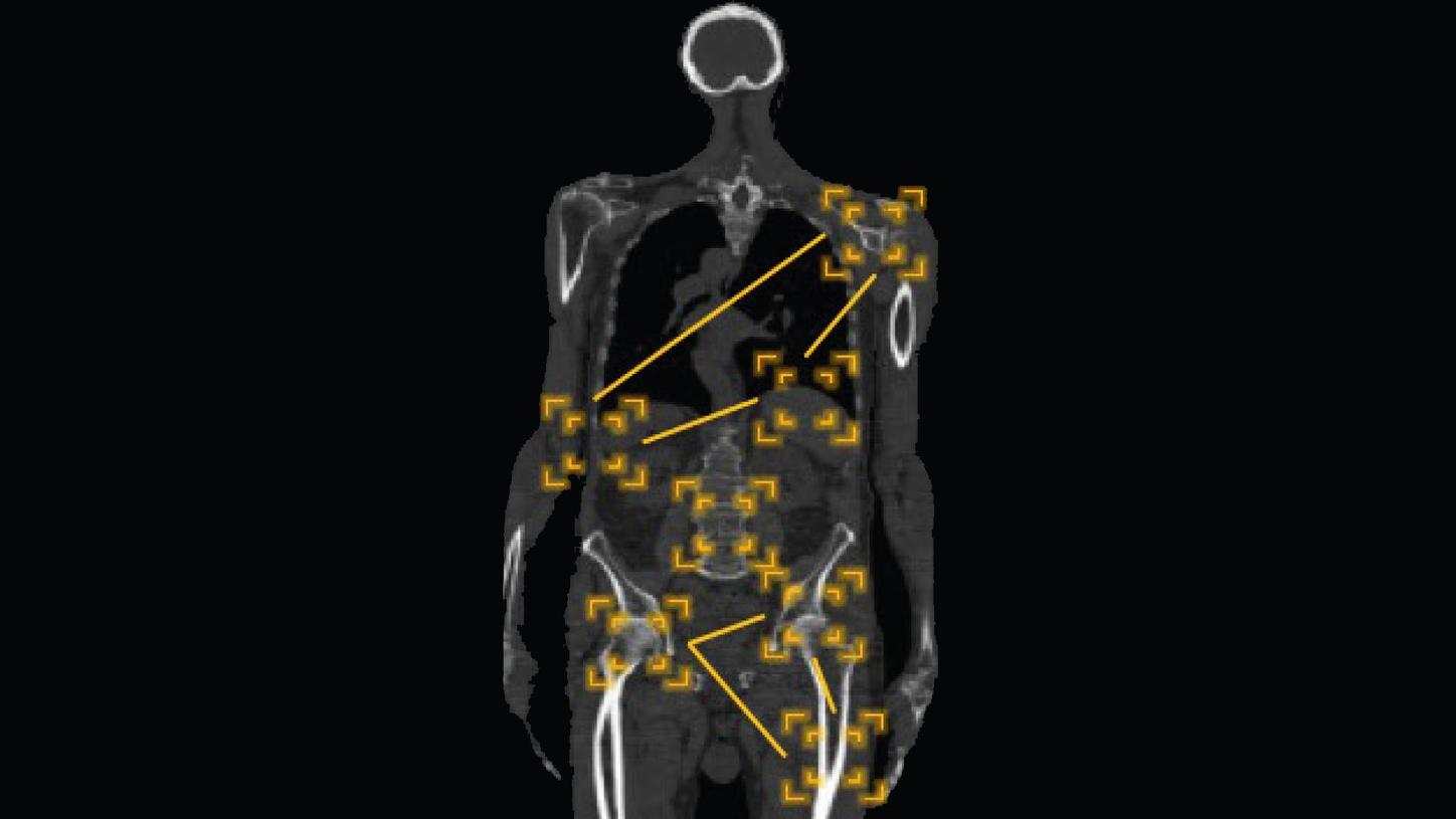
Minimize motion artifacts and optimize image quality with fast temporal resolution and the highest tube current of its class. Automated postprocessing provides comprehensive results for cardiac assessment.


Low-dose CT imaging
The secrets of low dose and excellent image quality
Integrate complex exams into daily practice: Equipped with premium technologies, SOMATOM go.Top delivers results you would not expect from a routine system. Find out more about our Stellar detector, High Power, Tin Filter, spectral imaging, iMAR, and SAFIRE.

Stellar detector
- Reduced image noise in every scan
- Excellent image quality at very low dose thanks to advanced iterative reconstruction SAFIRE1)
- Excellent and homogenous image quality, even in complex areas – due to an increased channel density and a new geometry
Benefits include:
- Up to 50% less dose to achieve equivalent image noise compared with conventional detectors
- Up to 45% fewer streak artifacts through regions of high attenuation (e.g., shoulder)2)

High Power, 10 kV Steps & CARE kV
- Highest tube current in its class: up to 825 mA even at 70 kV – thanks to the Athlon™ tube
- Better iodine contrast for sharper images, even in small distal vessels.
- Considerably reduced contrast media lets you scan more patients, deliver better patient care, and reduce examination costs
- CARE kV automatically tailors tube voltage to each patient and clinical indication
- Voltage levels can be adjusted at intervals of 10 kV for less dose and high contrast resolution

Tin Filter
- Unique technology from Siemens Healthineers
- Ultra-low dose levels by cutting out lower energies
- Optimized image quality at the interface between soft tissue and air
- Direct benefits, for example, in lung and colon imaging
- Reduced beam-hardening artifacts and improved image quality in bony structures
Spectral imaging with Dual Energy
- TwinSpiral Dual Energy is the next generation of spectral imaging in clinical routine
- Holistic solution with two Dual Energy modes prepares you for virtually all clinical questions
- TwinBeam Dual Energy acquires low- and high-kV data sets in a single scan – providing an unparalleled wealth of diagnostic information
- TwinSpiral Dual Energy provides both morphological and functional information for contrast media scans – by a new workflow concept of two scans integrated into a single acquisition
- Advanced automation with improved spectral separation due to the Tin Filter, better dose distribution, and the well-known GO technologies
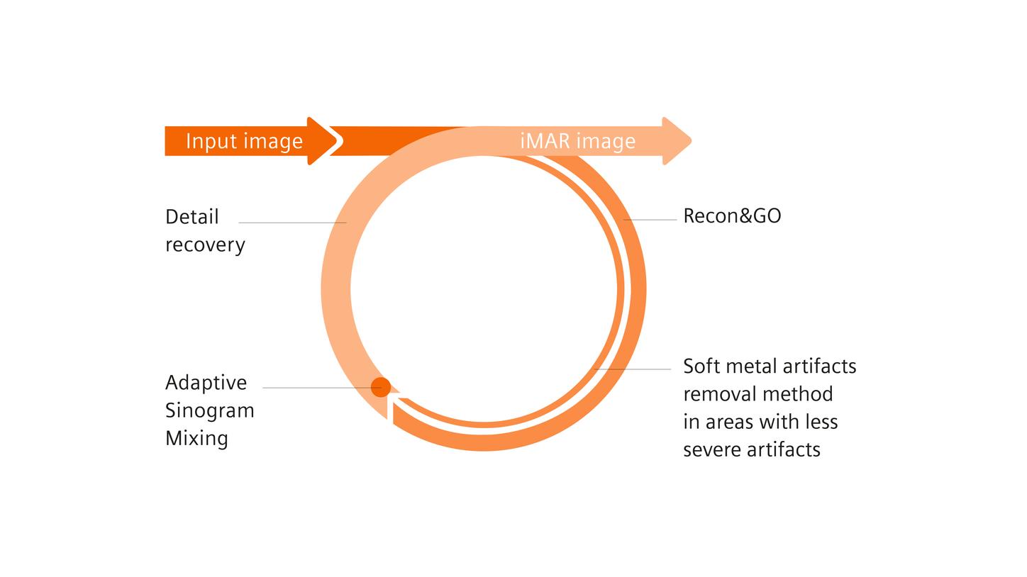
iMAR3)
- High-end algorithm reduces metal artifacts for better image quality without increase in dose
- Can handle a wide variety of metal implants for smoother, more efficient workflows
- Facilitates imaging of metal implants like dental fillings, pacemakers, and extremity implants

SAFIRE1)
- Iterative reconstruction algorithm
- Delivers excellent image quality at low dose
- Fast and simple to use
- Can be easily implemented into daily routine
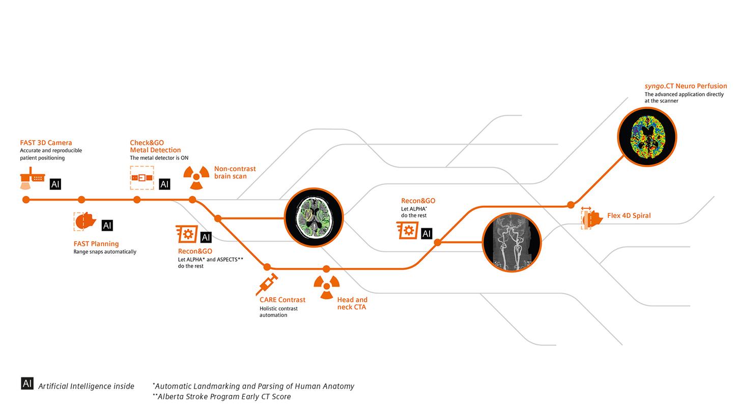
Smart CT technologies
Smart helpers that let you go for any pathway
The SOMATOM go.Top CT scanner features smart helpers to standardize and simplify your departmental processes – from patient setup to image distribution, archiving, and reading. Prevent repetitions. Skip routines. And dedicate your energy to patients and results.

myExam Companion
- Launches the era of intelligent imaging
- Helps users efficiently achieve reproducible results – by unlocking your modality’s full potential automatically
- Guides users through any procedure, so they can interact easily and naturally with both patient and technology
- No matter the user, patient or throughput, it helps generate consistently excellent results – and improve diagnostic confidence

AI-Rad Companion
- A family of vendor-neutral, multi-organ augmented reading solutions
- Takes over basic, repetitive tasks to support experienced staff in working at the top of their license
- Identifies and quantifies relevant anatomies and abnormalities
- Puts findings into a diagnostic context, for faster and more accurate diagnoses
Scan&GO
- Anticipates potential breathing artifacts
- Lets you control scans remotely and check images, right after the scan, on your tablet
- Up to three tablets can be used in perfect sync

Check&GO
- Intelligent algorithm based on big data
- Identifies potential errors with organ coverage and contrast media volume or distribution
- Identifies the presence of wearable metal objects like belts and necklaces
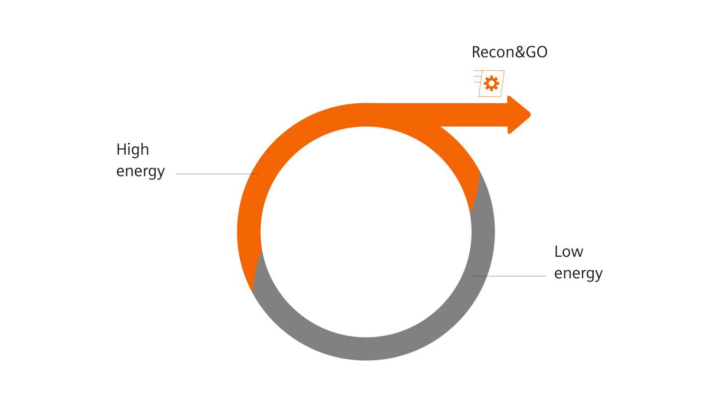
Recon&GO
- Delivers fast and standardized results irrespective of the operator
- AI recognizes patient landmarks and anatomies for automated postprocessing tasks
- Reduces repetitive workflow steps
- Even spectral imaging becomes routine with zero-click results
- Fully automated recon process for any organ – including all vascular views for contrast-enhanced CT reporting

CT View&GO
- Enables smooth reading in one workflow right at the scanner
- Advanced CAD algorithms and applications boost diagnostic confidence
- Makes communication within your department easy, since it automatically films and distributes images and results according to your settings
Guide&GO
- Brings our trendsetting Mobile Workflow to CT-guided interventions
- Dedicated high-end low-dose technologies
- Intuitive and flexible operation
- The only tablet-based solution in the market
iMAR – iterative metal artifact reduction – is designed to yield images with a reduced level of metal artifacts compared to conventional reconstruction if the underlying CT data is distorted by metal being present in the scanned object. The exact amount of metal artifact reduction and the corresponding improvement in image quality achievable depends on a number of factors, including composition and size of the metal part within the object, the patient size, anatomical location, and clinical practice. It is recommended to perform iMAR reconstruction in addition to conventional reconstruction.