- Home
- Medical Imaging
- Molecular Imaging
- PET/CT Scanners
- Biograph Vision
Biograph VisionPrecision-driven performance
Redefine PET/CT imaging with the precision-driven performance of Biograph Vision™1. With industry-leading technical specifications, Biograph Vision delivers next-level image quality that transforms accurate decision-making.2,3
![Siemens Healthineers Biograph Vision PET/CT scanner](https://marketing.webassets.siemens-healthineers.com/ae49b2acc7dbcfc7/3026969a70d6/v/0f314102981b/siemens-healthineers_mi_biograph_vision_pet-ct.jpg)
Benefits
![Biograph Vision PET/CT cardiac scan](https://marketing.webassets.siemens-healthineers.com/8294f0fe158027fe/66a2ee56d8a3/v/93747ca94863/siemens-healthineers_MI_biograph-vision-pet-ct-cardiology.png)
Biograph Vision’s groundbreaking technological performance redefines the limits of imaging. Featuring 3.2-mm lutetium oxyorthosilicate (LSO) crystals that are 100% covered by SiPMs2, Biograph Vision’s Optiso Ultra Dynamic Range (UDR) detector technology delivers high 48-mm3 volumetric resolution2 and 214-ps true time-of-flight (TOF) performance.3 Biograph Vision leverages the full potential of SiPM technology to reveal the bigger picture for accurate and confident decision-making.
![Biograph Vision PET/CT](https://marketing.webassets.siemens-healthineers.com/08ccf38c590e396c/a4d18790e96c/v/1d223ed5b161/siemens-healthineers_MI_biograph-vision-pet-ct-female-whole-body.png)
Unlock more clinical and research capabilities with Biograph Vision’s state-of-the-art detectability. Biograph Vision provides high image resolution and sensitivity for precise quantification that can positively impact health outcomes. Ultra Dynamic Range (UDR) provides outstanding performance for a wide spectrum of count rates, enabling a broad variety of radiotracers at optimal doses.
![Biograph Vision PET/CT radiation therapy](https://marketing.webassets.siemens-healthineers.com/95f89388685b1149/021acb6d0339/v/00129c79a122/siemens-healthineers_MI_biograph-vision-pet-ct-radiation-therapy.png)
Biograph Vision empowers users to reduce scan time and injected dose to boost productivity, avoid unnecessary exposure, and increase patient comfort with the market’s highest effective sensitivity3 at 100 cps/kBq. Enhance patient and user experience with intelligent imaging capabilities that drive greater throughput while providing more consistent and accurate results. No matter the user, the patient,5 or the procedure, Biograph Vision delivers exceptional outcomes.
![Biograph Vision PET/CT cardiac scan](https://marketing.webassets.siemens-healthineers.com/8294f0fe158027fe/66a2ee56d8a3/v/93747ca94863/siemens-healthineers_MI_biograph-vision-pet-ct-cardiology.png)
Biograph Vision’s groundbreaking technological performance redefines the limits of imaging. Featuring 3.2-mm lutetium oxyorthosilicate (LSO) crystals that are 100% covered by SiPMs2, Biograph Vision’s Optiso Ultra Dynamic Range (UDR) detector technology delivers high 48-mm3 volumetric resolution2 and 214-ps true time-of-flight (TOF) performance.3 Biograph Vision leverages the full potential of SiPM technology to reveal the bigger picture for accurate and confident decision-making.
Evidence
Case 1
02Case 2
04Case 3
04Case 4
04Case 5
02Case 6
03Case 7
03Case 8
03![Molecular Imaging Biograph Vision Case 3](https://marketing.webassets.siemens-healthineers.com/ca595880746ad7e0/eb8e9326084b/v/aced288697b4/siemens-healthineers-mi-biograph-case8-3.png)
Data courtesy of University Medical Center Groningen, Groningen, The Netherlands.
Case 8
Delineation of multiple skeletal, lymph node, liver, and soft tissue metastases with 18F FDG PET/CT
Multiple hypermetabolic metastases in the liver, mediastinal lymph nodes, vertebrae, and soft tissue sharply delineated in 18F FDG[a] PET/CT study on Biograph Vision reconstructed with high resolution at 440 x 440 matrix.
Examination protocol
Biograph Vision, 8.2 mCi (305 MBq) 18F FDG, 1 hour 41 minutes post-injection delay.
CT: 100 kV, 11 eff mAs. 32 x 1.2 mm collimation, PET: 7 beds 3 minutes/bed 440 x 440 matrix.
![Biograph Vision clinical image brain](https://marketing.webassets.siemens-healthineers.com/ec20c12681291eaf/439ed2971717/v/164086ff536d/siemens-healthineers-mi-biograph-clinical-gallery-brain.png)
Data courtesy of University Medical Center Groningen, Groningen, The Netherlands.
Case 1
Normal brain metabolism in a subject with suspected multi-system atrophy
Normal brain study with sharp delineation of functioning cortex and increased contrast between cortex and white matter.
Clinical detail
A 67-year-old male with progressive muscular stiffness, difficulty in swallowing, and autonomic dysfunctions with no response to levodopa therapy; undergoing evaluation for striatal multi-system atrophy. A Fludeoxyglucose F 18 Injection.[a] (18F FDG) PET/CT scan of the brain was performed on Biograph Vision. The study shows normal uptake in brain with excellent delineation of functioning cortex and sharp contrast between cortex and white matter. Additionally, there is sharp basal ganglial edge definition, especially the sharp margins and distinct separation of the head of caudate nucleus and putamen.
![Biography Vision clinical image brain](https://marketing.webassets.siemens-healthineers.com/562ce676f6f67e43/3c5ce79a6736/v/294076613e5b/siemens-healthineers-mi-biograph-Biograph-Vision-clinical-image-brain-2.png)
Data courtesy of University Medical Center Groningen, Groningen, The Netherlands.
Case 1
Normal brain metabolism in a subject with suspected multi-system atrophy
Normal brain study with sharp delineation of functioning cortex and increased contrast between cortex and white matter.
Examination protocol
Biograph Vision, 5.4 mCi (200 MBq)18F FDG, 51 minutes post-injection delay.
CT: 80 kV, 30 eff mAs. 128 x 0.6 mm collimation, PET: 15 minutes total scan time 440 x 440 matrix.
Data courtesy of University Medical Center Groningen, Groningen, The Netherlands.
Case 2
Visualization of vascular blood pool during whole-body dynamic PET/CT acquisition
Fluoro-dihydrotestosterone (18F FDHT)[b] PET/CT study in a patient with prostate cancer with biochemical relapse. Sequential whole-body dynamic FlowMotion™ acquisitions shortly after 18F FDHT administration demonstrates blood pool activity within heart and major vessels as well as tracer clearance through liver, kidneys, and intestine.
Clinical detail
A 73-year-old male with prostate cancer treated with brachytherapy presented with biochemical relapse. The patient underwent PET/CT study following injection of androgen receptor imaging agent Fluoro-dihydrotestosterone (18F-FDHT) in order to assess presence of androgen receptor positive recurrence. PET/CT was performed on Biograph Vision. Images showed tracer within cardiac and vascular blood pool as well as in the liver. Significant clearance of tracer through the biliary tract and gall bladder, kidneys, and intestines is visualized. No well defined focal uptake suggestive of androgen receptor-avid metastatic lesions is visualized, although primary prostate bed showed significant uptake on higher intensity images. Sharp delineation of vascular structures, especially cavernous and sigmoid sinuses as well as carotid and subclavian vessels reflect the high overall image quality and low noise level in the Biograph Vision images.
![Molecular Imaging Biograph Vision Case 2-2](https://marketing.webassets.siemens-healthineers.com/bfbed94c4d5bfcc6/fd8ffe8efa79/v/6db63b8c773d/siemens-healthineers-mi-biograph-case2-2.png)
Data courtesy of University Medical Center Groningen, Groningen, The Netherlands.
Case 2
Visualization of vascular blood pool during whole-body dynamic PET/CT acquisition
Fluoro-dihydrotestosterone (18F FDHT)[b] PET/CT study in a patient with prostate cancer with biochemical relapse. Sequential whole-body dynamic FlowMotion™ acquisitions shortly after 18F FDHT administration demonstrates blood pool activity within heart and major vessels as well as tracer clearance through liver, kidneys, and intestine.
Examination protocol
Biograph Vision, 7.5 mCi (200 MBq) 18F FDHT, 1 hour 35 minutes post-injection delay.
CT: 100 kV, 13 eff mAs. 32 x 1.2 mm collimation, PET: Dynamic acquisition 220 x 220 matrix.
![Molecular Imaging Biograph Vision Case 2-3](https://marketing.webassets.siemens-healthineers.com/52080741a863016e/d928c5074dd6/v/bd3c34a22106/siemens-healthineers-mi-biograph-case2-3.png)
Data courtesy of University Medical Center Groningen, Groningen, The Netherlands.
Case 2
Visualization of vascular blood pool during whole-body dynamic PET/CT acquisition
Fluoro-dihydrotestosterone (18F FDHT)[b] PET/CT study in a patient with prostate cancer with biochemical relapse. Sequential whole-body dynamic FlowMotion™ acquisitions shortly after 18 F FDHT administration demonstrates blood pool activity within heart and major vessels as well as tracer clearance through liver, kidneys, and intestine.
Examination protocol
Biograph Vision, 7.5 mCi (200 MBq) 18F FDHT, 1 hour 35 minutes post-injection delay.
CT: 100 kV, 13 eff mAs. 32 x 1.2 mm collimation, PET: Dynamic acquisition 220 x 220 matrix.
![Molecular Imaging Biograph Vision Case 2-4](https://marketing.webassets.siemens-healthineers.com/0853b9e4b83a4c30/d2736eec7f33/v/68425b9cdc27/siemens-healthineers-mi-biograph-case2-4.png)
Data courtesy of University Medical Center Groningen, Groningen, The Netherlands.
Case 2
Visualization of vascular blood pool during whole-body dynamic PET/CT acquisition
Fluoro-dihydrotestosterone (18F FDHT)[b] PET/CT study in a patient with prostate cancer with biochemical relapse. Sequential whole-body dynamic FlowMotion™ acquisitions shortly after 18F FDHT administration demonstrates blood pool activity within heart and major vessels as well as tracer clearance through liver, kidneys, and intestine.
Examination protocol
Biograph Vision, 7.5 mCi (200 MBq) 18F FDHT, 1 hour 35 minutes post-injection delay.
CT: 100 kV, 13 eff mAs. 32 x 1.2 mm collimation, PET: Dynamic acquisition 220 x 220 matrix.
![Molecular Imaging Biograph Vision Case Prostate](https://marketing.webassets.siemens-healthineers.com/b30788c6e45344ad/e7b58dcb5e9a/v/5c2ffc2c6148/siemens-healthineers-mi-biograph-clinical-gallery-prostate.png)
Data courtesy of University Medical Center Groningen, Groningen, The Netherlands.
Case 3
Visualization of 7 mm lymph node metastases on 68Ga-PSMA evaluation of primary prostate cancer
68Ga-PSMA[b] PET/CT in a patient with primary prostate cancer shows PSMA-avid primary tumor along with small PSMA-avid right external iliac lymph node metastases. Uptake in the kidneys, salivary gland, liver, intestine, and urinary bladder are within physiological limits.
Clinical detail
A 78-year-old male with primary prostate cancer underwent 68Ga-PSMA PET/CT initial staging. PET/CT was performed on Biograph Vision. The images showed high tracer uptake in kidneys, salivary glands, and bladder, which are at physiologically normal levels. The liver, spleen, and intestines also show lower but normal levels of uptake as well. There is also focal hypermetabolism in the prostate reflecting primary tumor and the small hypermetabolic right external iliac lymph node is suggestive of solitary nodal metastases. No other PSMA-avid metastatic lesions are visualized. Sharply delineated renal, salivary gland, bladder, and intestinal margins reflect the high overall image quality and low background noise level in the Biograph Vision images.
![Molecular Imaging Biograph Vision Case 3-2](https://marketing.webassets.siemens-healthineers.com/79a4b1934a4e6f17/8e0fb1fb8247/v/c2057c04c025/siemens-healthineers-mi-biograph-case3-2.png)
Data courtesy of University Medical Center Groningen, Groningen, The Netherlands.
Case 3
Visualization of 7 mm lymph node metastases on 68Ga-PSMA evaluation of primary prostate cancer
68Ga-PSMA[b] PET/CT in a patient with primary prostate cancer shows PSMA-avid primary tumor along with small PSMA-avid right external iliac lymph node metastases. Uptake in the kidneys, salivary gland, liver, intestine, and urinary bladder are within physiological limits.
Examination protocol
Biograph Vision, 2.7 mCi (100 MBq) 68Ga-PSMA, 1 hour 45 minutes post-injection delay.
CT: 100 kV, 16 eff mAs. 32 x 1.2 mm collimation, PET: 220 x 220 matrix.
![Molecular Imaging Biograph Vision Case 3-3](https://marketing.webassets.siemens-healthineers.com/161c3c4b98b56f89/fab3c1432a7f/v/6efec2c84778/siemens-healthineers-mi-biograph-case3-3.png)
Data courtesy of University Medical Center Groningen, Groningen, The Netherlands.
Case 3
Visualization of 7 mm lymph node metastases on 68Ga-PSMA evaluation of primary prostate cancer
Visualization of 7 mm lymph node metastases on 68Ga-PSMA[b] evaluation of primary prostate cancer
68Ga-PSMA PET/CT in a patient with primary prostate cancer shows PSMA-avid primary tumor along with small PSMA-avid right external iliac lymph node metastases. Uptake in the kidneys, salivary gland, liver, intestine, and urinary bladder are within physiological limits.
Examination protocol
Biograph Vision, 2.7 mCi (100 MBq) 68Ga-PSMA, 1 hour 45 minutes post-injection delay.
CT: 100 kV, 16 eff mAs. 32 x 1.2 mm collimation, PET: 220 x 220 matrix.
![Molecular Imaging Biograph Vision Case 3-4](https://marketing.webassets.siemens-healthineers.com/d6637cb111b5b8be/326887b0d3fb/v/8c83b9bca84c/siemens-healthineers-mi-biograph-case3-4.png)
Data courtesy of University Medical Center Groningen, Groningen, The Netherlands.
Case 3
Visualization of 7 mm lymph node metastases on 68Ga-PSMA evaluation of primary prostate cancer
Visualization of 7-mm lymph node metastases on 68Ga-PSMA[b] evaluation of primary prostate cancer
68Ga-PSMA PET/CT in a patient with primary prostate cancer shows PSMA-avid primary tumor along with small PSMA-avid right external iliac lymph node metastases. Uptake in the kidneys, salivary gland, liver, intestine, and urinary bladder are within physiological limits.
Examination protocol
Biograph Vision, 2.7 mCi (100 MBq) 68Ga-PSMA, 1 hour 45 minutes post-injection delay.
CT: 100 kV, 16 eff mAs. 32 x 1.2 mm collimation, PET: 220 x 220 matrix.
![Molecular Imaging Biograph Vision Case 4-1](https://marketing.webassets.siemens-healthineers.com/dd210c35332ee9ff/6b0551d2d416/v/2057893dcfb5/siemens-healthineers-mi-biograph-case4-1.png)
Data courtesy of University Medical Center Groningen, Groningen, The Netherlands.
Case 4
High contrast in pelvic nodal and hepatic metastases in a patient with operated ovarian carcinoma seen by 18F FDG PET/CT
Hypermetabolic right obturator and liver metastases visualized with high contrast on 18F FDG[a] PET/CT in a patient with operated ovarian carcinoma.
Clinical detail
A 49-year-old woman with a history of operated ovarian carcinoma with suspected recurrence underwent 18F FDG PET/CT for restaging. A PET/CT was performed on Biograph Vision. The images showed hypermetabolic metastases in the right obturator node, anterior pelvic wall above, and adjacent to the bladder as well as multiple small liver metastases in the posterior aspect of the right lobe (Segment VII) and in the caudate lobe. Sharp delineation of small liver lesions with high contrast compared to normal liver without respiratory gating reflect the overall high image quality and high lesion contrast to background in the Biograph Vision images.
![Molecular Imaging Biograph Vision Case 4-2](https://marketing.webassets.siemens-healthineers.com/4c74ef44ef2d2bd5/04c324e5f195/v/56073106dd71/siemens-healthineers-mi-biograph-case4-2.png)
Data courtesy of University Medical Center Groningen, Groningen, The Netherlands.
Case 4
High contrast in pelvic nodal and hepatic metastases in a patient with operated ovarian carcinoma seen by 18F FDG PET/CT
Hypermetabolic right obturator and liver metastases visualized with high contrast on 18F FDG[a] PET/CT in a patient with operated ovarian carcinoma.
Examination protocol
Biograph Vision, 7.5 mCi (280 MBq) 18 FDG, 1 hour 49 minutes post-injection delay.
CT: 140 kV, 16 eff mAs. 32 x 1.2 mm collimation, PET: 7 beds 3 minutes/bed 220 x 220 matrix.
![Molecular Imaging Biograph Vision Case 4-3](https://marketing.webassets.siemens-healthineers.com/64315b05c67fe29a/081c1a9ba275/v/fd440a9e5328/siemens-healthineers-mi-biograph-case4-3.png)
Data courtesy of University Medical Center Groningen, Groningen, The Netherlands.
Case 4
High contrast in pelvic nodal and hepatic metastases in a patient with operated ovarian carcinoma seen by 18F FDG PET/CT
Hypermetabolic right obturator and liver metastases visualized with high contrast on 18F FDG[a] PET/CT in a patient with operated ovarian carcinoma.
Examination protocol
Biograph Vision, 7.5 mCi (280 MBq) 18 FDG, 1 hour 49 minutes post-injection delay.
CT: 140 kV, 16 eff mAs. 32 x 1.2 mm collimation, PET: 7 beds 3 minutes/bed 220 x 220 matrix.
![Molecular Imaging Biograph Vision Case 4-4](https://marketing.webassets.siemens-healthineers.com/9613f468c4bf7329/4b1efd8ac9a8/v/987e71a40796/siemens-healthineers-mi-biograph-case4-4.png)
Data courtesy of University Medical Center Groningen, Groningen, The Netherlands.
Case 4
High contrast in pelvic nodal and hepatic metastases in a patient with operated ovarian carcinoma seen by 18F FDG PET/CT
Hypermetabolic right obturator and liver metastases visualized with high contrast on 18F FDG[a] PET/CT in a patient with operated ovarian carcinoma.
Examination protocol
Biograph Vision, 7.5 mCi (280 MBq) 18 FDG, 1 hour 49 minutes post-injection delay.
CT: 140 kV, 16 eff mAs. 32 x 1.2 mm collimation, PET: 7 beds 3 minutes/bed 220 x 220 matrix.
![Molecular Imaging Biograph Vision Case 5-1](https://marketing.webassets.siemens-healthineers.com/2b5ad8f3b15c74bd/08f8f5aac336/v/d8913c43279e/siemens-healthineers-mi-biograph-case5-1.png)
Data courtesy of University Medical Center Groningen, Groningen, The Netherlands.
Case 5
Small liver metastases in a patient with operated colorectal carcinoma detected with 18F FDG PET/CT
Small hypermetabolic foci in the right lobe of liver close to the diaphragm visualized with high contrast and minimal motion related blurring with 18F FDG[a] PET/CT.
Clinical detail
A 69-year-old male with a history of operated colorectal carcinoma with suspected recurrence underwent 18F FDG PET/CT for restaging. A PET/CT was performed on Biograph Vision. The images showed sharp delineation of small hypermetabolic metastasis in the right lobe of liver near the dome of diaphragm (Segment VII). Intense uptake in the abdominal wall is suggestive of metastatic deposit. Mildly increased uptake in mediastinal lymph nodes are possibly secondary to inflammation. High contrast of small liver metastases without significant motion related blurring even without respiratory gating reflects high overall image quality and increased lesion contrast to background in the Biograph Vision images.
![Molecular Imaging Biograph Vision Case 5-2](https://marketing.webassets.siemens-healthineers.com/734b69d265f92f4f/d08dc06c3de6/v/13483adc6ea8/siemens-healthineers-mi-biograph-case5-2.png)
Data courtesy of University Medical Center Groningen, Groningen, The Netherlands.
Case 5
Small liver metastases in a patient with operated colorectal carcinoma detected with 18F FDG PET/CT
Small hypermetabolic foci in the right lobe of liver close to the diaphragm visualized with high contrast and minimal motion related blurring with 18F FDG[a] PET/CT.
Examination protocol
Biograph Vision, 7.3 mCi (270 MBq) 18F FDG, 1 hour 50 minutes post-injection delay. CT: 100 kV, 10 eff mAs. 32 x 1.2 mm collimation, PET: 7 beds 3 minutes/bed 440 x 440 matrix.
![Molecular Imaging Biograph Vision Case](https://marketing.webassets.siemens-healthineers.com/6d1fc4f7b7b3397e/34d22a553bdc/v/d8dfffee8a6c/siemens-healthineers-mi-biograph-case6-1.png)
Data courtesy of University Medical Center Groningen, Groningen, The Netherlands.
Case 6
18F-Fluroestradiol PET/CT in a patient with breast carcinoma
Sharp visualization of physiological uptake in biliary tract, gall bladder, liver, and intestines in a patient with breast carcinoma without estrogen receptor positive metastases undergoing PET/CT imaging with 18F-Fluoroestradiol (FES).[b]
Clinical detail
A 77-year-old female with a history of breast carcinoma treated with left mastectomy and chemo-radiotherapy presented with pain in the thoracic wall along with pleural effusion. The patient underwent 18F Fluoroestradiol (FES) PET/CT to assess presence of estrogen receptor (ER) positive metastases. A PET/CT was performed on Biograph Vision. High resolution PET images show faint uptake in the left pleura suggestive of ER positive pleural thickening. The PET/CT images also sharply delineates physiological uptake in the gall bladder and biliary tract and intestine reflecting hepatobiliary clearance of tracer. Sharp definition of biliary tree with high contrast compared to background even at 440 x 440 matrix reconstructed resolution reflect the high overall image quality of Biograph Vision images.
![Molecular Imaging Biograph Vision Case 6-2](https://marketing.webassets.siemens-healthineers.com/bcc114cc97421138/2ef23b04cb94/v/88f3dcc61c67/siemens-healthineers-mi-biograph-case6-2.png)
Data courtesy of University Medical Center Groningen, Groningen, The Netherlands.
Case 6
18F-Fluroestradiol PET/CT in a patient with breast carcinoma
Sharp visualization of physiological uptake in biliary tract, gall bladder, liver, and intestines in a patient with breast carcinoma without estrogen receptor positive metastases undergoing PET/CT imaging with 18F-Fluoroestradiol (FES).[b]
Examination protocol
Biograph Vision, 5.2 mCi (195 MBq) 18F FES, 1 hour 29 minutes post-injection delay.
CT: 100 kV, 13 eff mAs. 32 x 1.2 mm collimation, PET: 7 beds 3 minutes/bed 440 x 440 matrix.
![Molecular Imaging Biograph Vision Case 6-3](https://marketing.webassets.siemens-healthineers.com/bd57eb2d1d391c6e/0638568b0f07/v/001f5f62eed9/siemens-healthineers-mi-biograph-case6-3.png)
Data courtesy of University Medical Center Groningen, Groningen, The Netherlands.
Case 6
18F-Fluroestradiol PET/CT in a patient with breast carcinoma
Sharp visualization of physiological uptake in biliary tract, gall bladder, liver, and intestines in a patient with breast carcinoma without estrogen receptor positive metastases undergoing PET/CT imaging with 18F-Fluoroestradiol (FES).[b]
Examination protocol
Biograph Vision, 5.2 mCi (195 MBq) 18F FES, 1 hour 29 minutes post-injection delay.
CT: 100 kV, 13 eff mAs. 32 x 1.2 mm collimation, PET: 7 beds 3 minutes/bed 440 x 440 matrix.
![Molecular Imaging Biograph Vision Case 7-1](https://marketing.webassets.siemens-healthineers.com/937c1066ea180ec8/c168693140b7/v/2f47923299be/siemens-healthineers-mi-biograph-case7-1.png)
Data courtesy of University Medical Center Groningen, Groningen, The Netherlands.
Case 7
Sharp delineation of physiological uptake in vascular walls, marrow, and intestine with 18F FDG PET/CT
Physiological uptake in aortic walls, carotid and subclavian vessels as well as normal marrow uptake in the vertebrae, ribs and pelvis sharply delineated in 18F FDG[a] PET/CT study on Biograph Vision reconstructed with high resolution at 440 x 440 matrix.
Clinical detail
A 79-year-old male with unknown clinical history. A PET/CT was performed on Biograph Vision. High resolution PET images show sharp delineation of physiological uptake in the aortic walls, atrial walls, carotid, and subclavian vessels. Normal marrow uptake within vertebrae, ribs, and pelvis are well visualized. Physiological uptake in the gastric mucosa and intestines, with note made of sharply delineated descending colon. Sharp delineation of normal structures like rib and vessels with low background uptake reflect high overall image quality even at 440x440 matrix reconstructed resolution of Biograph Vision images.
![Molecular Imaging Biograph Vision Case 7-2](https://marketing.webassets.siemens-healthineers.com/2e4f58ae1600b1a4/bf480fb2ba40/v/bc0f79f1963d/siemens-healthineers-mi-biograph-case7-2.png)
Data courtesy of University Medical Center Groningen, Groningen, The Netherlands.
Case 7
Sharp delineation of physiological uptake in vascular walls, marrow, and intestine with 18F FDG PET/CT
Physiological uptake in aortic walls, carotid and subclavian vessels as well as normal marrow uptake in the vertebrae, ribs and pelvis sharply delineated in 18F FDG[a] PET/CT study on Biograph Vision reconstructed with high resolution at 440 x 440 matrix.
Examination protocol
Biograph Vision, 8.2 mCi (305 MBq) 18F FDG, 1 hour 41 minutes post-injection delay.
CT: 100 kV, 11 eff mAs. 32 x 1.2 mm collimation, PET: 7 beds 3 minutes/bed 440 x 440 matrix.
![Molecular Imaging Biograph Vision Case 7-3](https://marketing.webassets.siemens-healthineers.com/13261656204a184a/0b2d1da5188a/v/7aab413f8f56/siemens-healthineers-mi-biograph-case7-3.png)
Data courtesy of University Medical Center Groningen, Groningen, The Netherlands.
Case 7
Sharp delineation of physiological uptake in vascular walls, marrow, and intestine with 18F FDG PET/CT
Sharp delineation of physiological uptake in vascular walls, marrow, and intestine with 18F FDG[a] PET/CT
Physiological uptake in aortic walls, carotid and subclavian vessels as well as normal marrow uptake in the vertebrae, ribs and pelvis sharply delineated in 18F FDG PET/CT study on Biograph Vision reconstructed with high resolution at 440 x 440 matrix.
Examination protocol
Biograph Vision, 8.2 mCi (305 MBq) 18F FDG, 1 hour 41 minutes post-injection delay.
CT: 100 kV, 11 eff mAs. 32 x 1.2 mm collimation, PET: 7 beds 3 minutes/bed 440 x 440 matrix.
![Molecular Imaging Biograph Vision Case 8-1](https://marketing.webassets.siemens-healthineers.com/0c02227e19a6a46b/9ad3f70af631/v/0483afba9148/siemens-healthineers-mi-biograph-case8-1.png)
Data courtesy of University Medical Center Groningen, Groningen, The Netherlands.
Case 8
Delineation of multiple skeletal, lymph node, liver, and soft tissue metastases with 18F FDG PET/CT
Multiple hypermetabolic metastases in the liver, mediastinal lymph nodes, vertebrae, and soft tissue sharply delineated in 18F FDG[a] PET/CT study on Biograph Vision reconstructed with high resolution at 440 x 440 matrix.
Clinical detail
70-year-old male. Though clinical history unknown, there are obviously multiple metastases as shown on PET/CT performed on the Biograph Vision. High resolution PET images show multiple hypermetabolic metastases in the liver, mediastinal lymph nodes, vertebrae and soft tissue. Sharp delineation with high lesion contrast of small metastases in the vertebrae, pelvis, mediastinal nodes and soft tissue especially in the left thigh reflect the high image quality even at 440x440 matrix reconstructed resolution of the Biograph Vision images.
![Molecular Imaging Biograph Vision Case 8-2](https://marketing.webassets.siemens-healthineers.com/c458d5a34e515527/e0dacdf63b5d/v/b82eb8de9f54/siemens-healthineers-mi-biograph-case8-2.png)
Data courtesy of University Medical Center Groningen, Groningen, The Netherlands.
Case 8
Delineation of multiple skeletal, lymph node, liver, and soft tissue metastases with 18F FDG PET/CT
Multiple hypermetabolic metastases in the liver, mediastinal lymph nodes, vertebrae, and soft tissue sharply delineated in 18F FDG[a] PET/CT study on Biograph Vision reconstructed with high resolution at 440 x 440 matrix.
Examination protocol
Biograph Vision, 8.2 mCi (305 MBq) 18F FDG, 1 hour 41 minutes post-injection delay.
CT: 100 kV, 11 eff mAs. 32 x 1.2 mm collimation, PET: 7 beds 3 minutes/bed 440 x 440 matrix.
![Molecular Imaging Biograph Vision Case 3](https://marketing.webassets.siemens-healthineers.com/ca595880746ad7e0/eb8e9326084b/v/aced288697b4/siemens-healthineers-mi-biograph-case8-3.png)
Data courtesy of University Medical Center Groningen, Groningen, The Netherlands.
Case 8
Delineation of multiple skeletal, lymph node, liver, and soft tissue metastases with 18F FDG PET/CT
Multiple hypermetabolic metastases in the liver, mediastinal lymph nodes, vertebrae, and soft tissue sharply delineated in 18F FDG[a] PET/CT study on Biograph Vision reconstructed with high resolution at 440 x 440 matrix.
Examination protocol
Biograph Vision, 8.2 mCi (305 MBq) 18F FDG, 1 hour 41 minutes post-injection delay.
CT: 100 kV, 11 eff mAs. 32 x 1.2 mm collimation, PET: 7 beds 3 minutes/bed 440 x 440 matrix.
![Biograph Vision clinical image brain](https://marketing.webassets.siemens-healthineers.com/ec20c12681291eaf/439ed2971717/v/164086ff536d/siemens-healthineers-mi-biograph-clinical-gallery-brain.png)
![Biography Vision clinical image brain](https://marketing.webassets.siemens-healthineers.com/562ce676f6f67e43/3c5ce79a6736/v/294076613e5b/siemens-healthineers-mi-biograph-Biograph-Vision-clinical-image-brain-2.png)
![Molecular Imaging Biograph Vision Case 2-2](https://marketing.webassets.siemens-healthineers.com/bfbed94c4d5bfcc6/fd8ffe8efa79/v/6db63b8c773d/siemens-healthineers-mi-biograph-case2-2.png)
![Molecular Imaging Biograph Vision Case 2-3](https://marketing.webassets.siemens-healthineers.com/52080741a863016e/d928c5074dd6/v/bd3c34a22106/siemens-healthineers-mi-biograph-case2-3.png)
![Molecular Imaging Biograph Vision Case 2-4](https://marketing.webassets.siemens-healthineers.com/0853b9e4b83a4c30/d2736eec7f33/v/68425b9cdc27/siemens-healthineers-mi-biograph-case2-4.png)
![Molecular Imaging Biograph Vision Case Prostate](https://marketing.webassets.siemens-healthineers.com/b30788c6e45344ad/e7b58dcb5e9a/v/5c2ffc2c6148/siemens-healthineers-mi-biograph-clinical-gallery-prostate.png)
![Molecular Imaging Biograph Vision Case 3-2](https://marketing.webassets.siemens-healthineers.com/79a4b1934a4e6f17/8e0fb1fb8247/v/c2057c04c025/siemens-healthineers-mi-biograph-case3-2.png)
![Molecular Imaging Biograph Vision Case 3-3](https://marketing.webassets.siemens-healthineers.com/161c3c4b98b56f89/fab3c1432a7f/v/6efec2c84778/siemens-healthineers-mi-biograph-case3-3.png)
![Molecular Imaging Biograph Vision Case 3-4](https://marketing.webassets.siemens-healthineers.com/d6637cb111b5b8be/326887b0d3fb/v/8c83b9bca84c/siemens-healthineers-mi-biograph-case3-4.png)
![Molecular Imaging Biograph Vision Case 4-1](https://marketing.webassets.siemens-healthineers.com/dd210c35332ee9ff/6b0551d2d416/v/2057893dcfb5/siemens-healthineers-mi-biograph-case4-1.png)
![Molecular Imaging Biograph Vision Case 4-2](https://marketing.webassets.siemens-healthineers.com/4c74ef44ef2d2bd5/04c324e5f195/v/56073106dd71/siemens-healthineers-mi-biograph-case4-2.png)
![Molecular Imaging Biograph Vision Case 4-3](https://marketing.webassets.siemens-healthineers.com/64315b05c67fe29a/081c1a9ba275/v/fd440a9e5328/siemens-healthineers-mi-biograph-case4-3.png)
![Molecular Imaging Biograph Vision Case 4-4](https://marketing.webassets.siemens-healthineers.com/9613f468c4bf7329/4b1efd8ac9a8/v/987e71a40796/siemens-healthineers-mi-biograph-case4-4.png)
![Molecular Imaging Biograph Vision Case 5-1](https://marketing.webassets.siemens-healthineers.com/2b5ad8f3b15c74bd/08f8f5aac336/v/d8913c43279e/siemens-healthineers-mi-biograph-case5-1.png)
![Molecular Imaging Biograph Vision Case 5-2](https://marketing.webassets.siemens-healthineers.com/734b69d265f92f4f/d08dc06c3de6/v/13483adc6ea8/siemens-healthineers-mi-biograph-case5-2.png)
![Molecular Imaging Biograph Vision Case](https://marketing.webassets.siemens-healthineers.com/6d1fc4f7b7b3397e/34d22a553bdc/v/d8dfffee8a6c/siemens-healthineers-mi-biograph-case6-1.png)
![Molecular Imaging Biograph Vision Case 6-2](https://marketing.webassets.siemens-healthineers.com/bcc114cc97421138/2ef23b04cb94/v/88f3dcc61c67/siemens-healthineers-mi-biograph-case6-2.png)
![Molecular Imaging Biograph Vision Case 6-3](https://marketing.webassets.siemens-healthineers.com/bd57eb2d1d391c6e/0638568b0f07/v/001f5f62eed9/siemens-healthineers-mi-biograph-case6-3.png)
![Molecular Imaging Biograph Vision Case 7-1](https://marketing.webassets.siemens-healthineers.com/937c1066ea180ec8/c168693140b7/v/2f47923299be/siemens-healthineers-mi-biograph-case7-1.png)
![Molecular Imaging Biograph Vision Case 7-2](https://marketing.webassets.siemens-healthineers.com/2e4f58ae1600b1a4/bf480fb2ba40/v/bc0f79f1963d/siemens-healthineers-mi-biograph-case7-2.png)
![Molecular Imaging Biograph Vision Case 7-3](https://marketing.webassets.siemens-healthineers.com/13261656204a184a/0b2d1da5188a/v/7aab413f8f56/siemens-healthineers-mi-biograph-case7-3.png)
![Molecular Imaging Biograph Vision Case 8-1](https://marketing.webassets.siemens-healthineers.com/0c02227e19a6a46b/9ad3f70af631/v/0483afba9148/siemens-healthineers-mi-biograph-case8-1.png)
![Molecular Imaging Biograph Vision Case 8-2](https://marketing.webassets.siemens-healthineers.com/c458d5a34e515527/e0dacdf63b5d/v/b82eb8de9f54/siemens-healthineers-mi-biograph-case8-2.png)
![Molecular Imaging Biograph Vision Case 3](https://marketing.webassets.siemens-healthineers.com/ca595880746ad7e0/eb8e9326084b/v/aced288697b4/siemens-healthineers-mi-biograph-case8-3.png)
Click plus (+) to find out our features
![Siemens Healthineers Biograph Vision](https://marketing.webassets.siemens-healthineers.com/9c2557b4355d867c/eb1824d3f890/v/78789e9f9007/siemens-healthineers_mi_biograph_vision_pet_ct_scanner_hotspot_image.jpg)
Exclusive bed design and wide bore
3.2-mm LSO crystals
Optiso UDR detector
Whole-body dynamic imaging
QualityGuard™CT capability
AIDAN Platform
Exclusive bed design and wide bore
Zero differential deflection for accurate attenuation correction and TG-66 compliant along with a 78-cm bore to support bariatric imaging and easier positioning of radiation oncology devices.
![Transcend digital with the Optiso UDR detector](https://marketing.webassets.siemens-healthineers.com/1800000004883487/ff69cda40da0/v/2674b0616030/siemens-healthineers_mi_optiso_udr_detector.jpg)
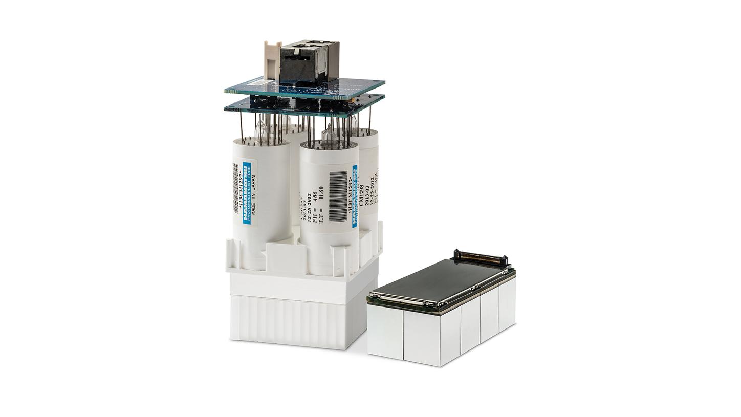
Take a look inside Biograph Vision - Transcend digital with the Optiso UDR detector
Optiso UDR’s proprietary 3.2-mm LSO crystals move silicon photomultiplier (SiPM) technology beyond digital to a new level of precision to help you detect small lesions, devise accurate treatment strategies, and achieve optimal performance in a wide range of count rates. See how the Optiso UDR PET detector advances PET/CT beyond digital.
![artificial intelligence application in nuclear medicine - AIDAN Intelligent imaging platform for PET CT scan machiness.](https://marketing.webassets.siemens-healthineers.com/f1304ad81d524d39/a151b242274b/v/b514954759b6/siemens-healthineers_mi_pet_ct_aidan_artificial_intelligence.png)
AIDAN Platform
Our intelligent imaging platform for Biograph™ PET/CT scanners that supports the demanding processing power of AI-based solutions.
Customer voices
"…by improving the spatial resolution…you have less partial volume effect, so you get sharper images and more accurate quantification."
![<p>Ronald Boellaard, PhD</p>](https://marketing.webassets.siemens-healthineers.com/1800000006199044/e74c33ed5ad8/v/494b3b18e68f/siemens-healthineers_mi_biograph_vision_quote_ronald_boellard.jpg)
Ronald Boellaard, PhD
Groningen, The Netherlands
"We have now already, as compared to the older system, reduced the activity we inject… Now it's probably 30% faster with about 30% less dose which is something very acceptable."
![John Prior, PhD, MD, FEBNM Lausanne, Switzerland](https://marketing.webassets.siemens-healthineers.com/1800000005821190/49a72022e5d5/v/b00466660d99/siemens-healthineers_mi_biograph_vision_quote_john_prior_switzerland.jpg)
John O. Prior, PhD, MD FEBNM
Lausanne, Switzerland
"Being more quantitative, our reproducibility can be that much better, and it may matter when we’re trying to do a repeat scan early on in a therapy and decide what to do."
![Richard E. Carson, PhD](https://marketing.webassets.siemens-healthineers.com/1800000006199045/7927cb48dedc/v/109894caec22/siemens-healthineers_mi_biograph_vision_quote_richard_carson.jpg)
New Haven, CT, USA
Biograph Vision.X
Biograph Vision.X PET/CT with exclusive, high-performance detector technology
Featured at Society of Nuclear Medicine and Molecular Imaging (SNMMI) 2023 annual meeting in Chicago, IL, USA, the new Biograph Vision.X™1 PET/CT builds on the proven performance of Biograph Vision and delivers an increased time of flight (TOF) of 178 ps—the industry’s fastest TOF.4
![Biograph Vision.X detector](https://marketing.webassets.siemens-healthineers.com/248dd1ac5effcbf2/fd2058ec0224/v/3ce54e39fb11/siemens-healthineers_mi_pet-ct_Biograph-VisionX_detector.png)
Biograph Vision.X builds on the proven performance of Biograph Vision and delivers:
- time of flight (TOF) performance of 178 picoseconds (ps)—the industry’s fastest TOF4
- outstanding performance gain up to 20%,5 which in turn can translate to faster scans as well as lower injected dose and radiotracer costs
- In-field upgradeability
Cutting-edge technology
Biograph Vision.X leverages our in-house production across the entire “imaging chain” and demonstrates our commitment to deliver cutting-edge PET/CT technology to our customers. Consider the power of each component along the imaging chain to deliver a better image.
Faster TOF
Biograph Vision.X with faster TOF reduces noise.
Upgradable
Biograph Vision.X is designed to be an in-field upgrade for installed Biograph Vision users, allowing them to extend the performance of their current system and offer next-level precision imaging.
"The Biograph Vision.X PET/CT scanner builds on the strong foundation of our Biograph Vision system and demonstrates our ongoing commitment to being the industry leader in time of flight for improved lesion detection and increased anatomical detail."
James Williams,
PhD Head of Molecular Imaging
Siemens Healthineers
Watch expert views on Biograph Vision
Accuracy to reveal the bigger picture
See smaller lesions, with better contrast. Hear what the experts say about the accuracy of Biograph Vision.
Performance to maximize efficiency
Market-leading time of flight and sensitivity2,7 mean lower patient exposure and reduced tracer cost.3 Learn what the performance of Biograph Vision can do for your patients.
Reproducibility to understand disease progression better
Absolute quantification and improved resolution and effective sensitivity3 enable you to evaluate treatment effectiveness earlier. Listen to the experts tell us how this affects their practice.
Watch our videos showing Biograph Vision become a reality
Making vision a reality
Watch the full-length documentary video on how Biograph Vision became a reality.
The passion behind the technology
Listen to first hand accounts from the talented men and women that worked on bringing Biograph Vision to reality.
Biograph Vision - Features
Learn more about how Biograph Vision goes beyond digital.
Technical Details
Bore diameter
78 cm
Tunnel length
136 cm
Table capacity
227 kg (500 lb)
Generator power
80 kW (100 kW optional)
Rotation times
0.33, 0.306, 0.286 s
Tube voltages
70, 80, 100, 120, 140 kV
Iterative reconstruction
SAFIRE6
Metal artifact reduction
iMAR6
Slices
64, 128
Axial field of view
26.3 cm
Crystal size
3.2 x 3.2 x 20 mm
SiPM coverage of crystal array
100%
Effective sensitivity
100 cps/kBq
Effective peak NEC rate
1,789 kcps ≤ 30 kBq/cc
Time of flight performance
214 ps
Related Topics
Downloads
Learn more about Biograph Vision
Based on patient weight limit of 227 kg (500 lb).
Based on competitive literature available at time of publication. Data on file.