- Home
- Medical Imaging
- Molecular Imaging
- Symbia Intevo
Symbia IntevoSPECT/CT that improves your image
Discover expanded clinical reach and a new standard of care. Symbia Intevo™ combines the improved image quality and localization of CT with proven SPECT capabilities, resulting in hybrid SPECT/CT technology that will distinguish your facility.

Benefits

Symbia Intevo offers high SPECT sensitivity1 and reconstructed resolution, along with high-performance CT, standard on every system. The result: confidence in knowing that every exam is performed with highest image quality.1
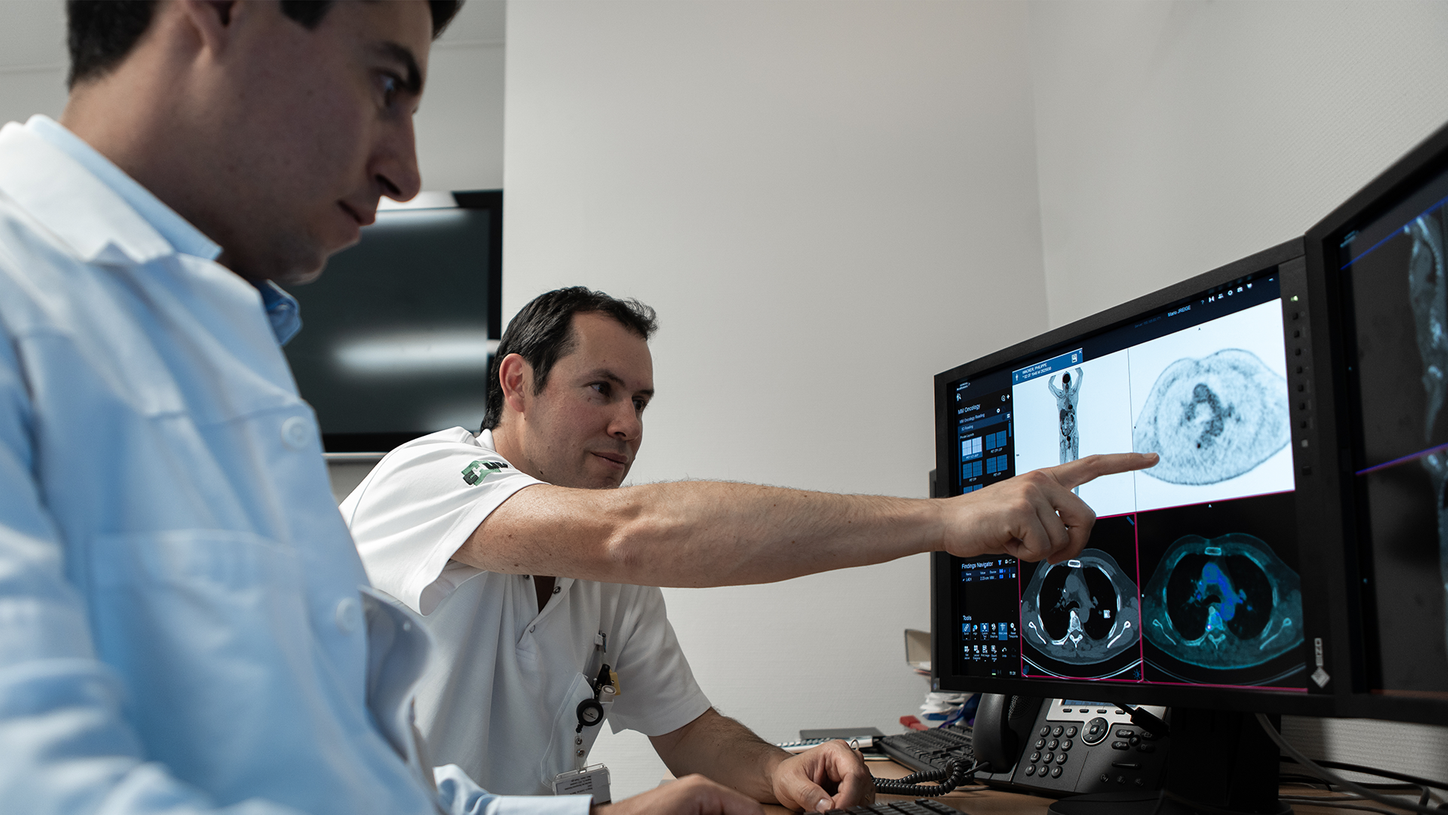
Symbia Intevo SPECT/CT imaging provides a more complete picture of your patient's condition in a single examination, allowing you to quickly make decisions that result in successful treatment strategies and a more satisfying experience.

Expand the scope of your imaging services with Symbia Intevo's advanced SPECT and CT technologies—and further distinguish your facility among referring physicians, patients, and the medical community through a reputation for providing quick, meaningful results.

Symbia Intevo offers high SPECT sensitivity1 and reconstructed resolution, along with high-performance CT, standard on every system. The result: confidence in knowing that every exam is performed with highest image quality.1
Evidence

Data courtesy of Universitat Wurzburg, Wurzburg, Germany.
SPECT/CT helps evaluate for inducible ischemia in a patient with a history of coronary disease
A SPECT/CT myocardial perfusion scan showed no sign of significant stress-induced ischemia or transmural scars, nor inferior and posterior wall attenuation with CTAC. The normal ejection fraction (EF) was 78%.
Data courtesy on file.
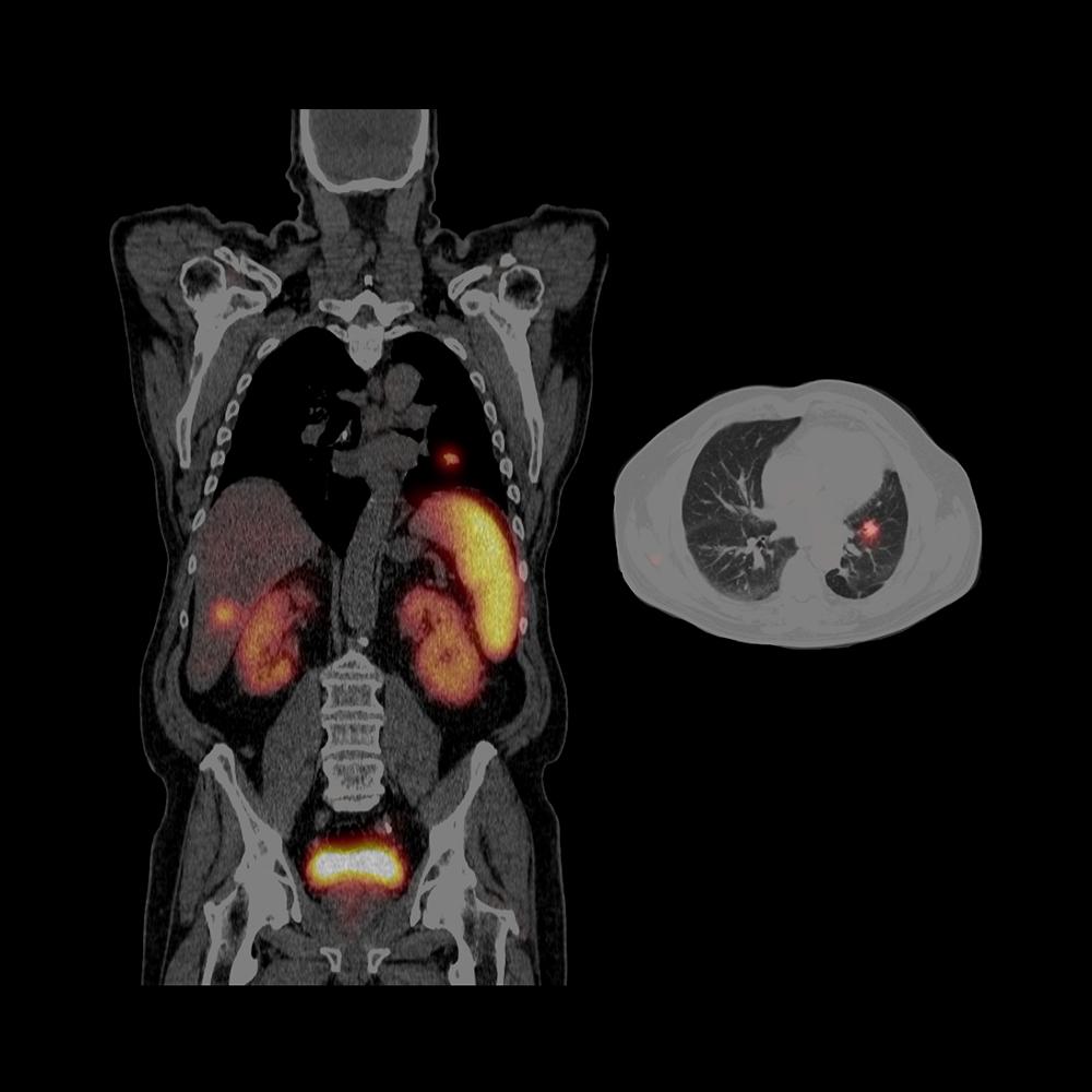
SPECT/CT assists in accurate localization of neuroendocrine tumor metastases
SPECT/CT localizes multiple tracer-avid somatostatin receptor-rich hepatic metastases, thereby helping in accurate localization of lesions and differentiation of lesion from physiological uptake.
Data courtesy of Holy Cross Hospital, Fort Lauderdale, Florida, USA.
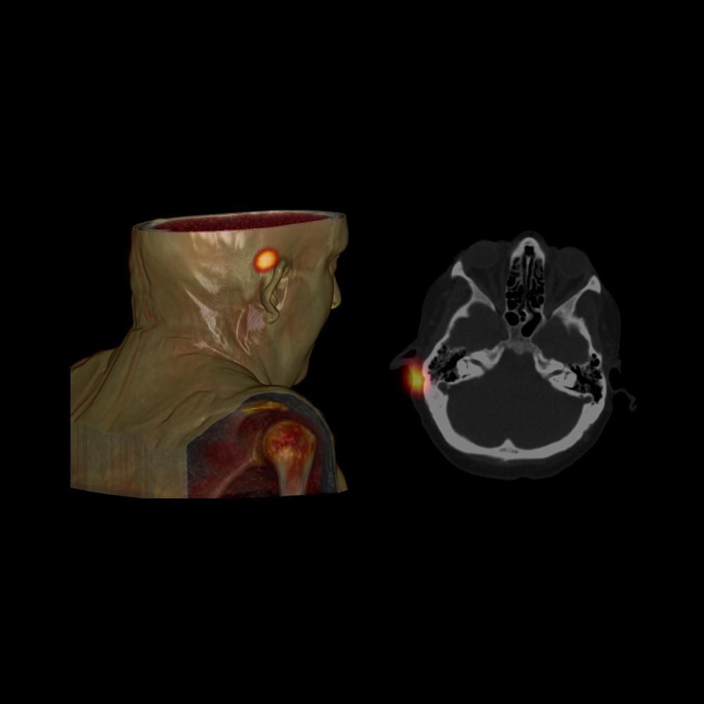
SPECT/CT localizes peri-auricular lymph sentinel node to aid in precise surgical removal
SPECT/CT imaging precisely defines a small peri-auricular sentinel lymph node due to accurate coregistration of anatomical information from the CT, with focal tracer uptake in the sentinel node seen on SPECT, allowing for precise surgical removal.

Data courtesy on file.
SPECT/CT accurately defines position of focal uptake to aid in localization of parathyroid adenoma
SPECT/CT-enabled localization of exact anatomical position of the parathyroid adenoma, which helped guide precise surgical exploration.

Data courtesy of Holy Cross Hospital, Fort Lauderdale, Florida, USA.
SPECT/CT helps characterize liver lesions with correlation of anatomical and functional information
CT shows focal hypodense liver lesions with absence of tracer uptake on SPECT. Correlation of anatomical information of liver lesions by CT and hepatocyte function by SPECT helps characterize the liver cysts.

Data courtesy of Centre Hospitalier Universitaire de Brest, Brest, France.
99mTc xSPECT Quant enables robust and reliable measurement of therapeutic effectiveness
xSPECT Quant™ across multiple time points quantifies the decrease of metabolic activity in the bone in a patient with metastases from prostate cancer. In addition, the uptake measured during anti-androgen therapy at the three time points correlated with a decline of prostate-specific antigen (PSA).

Data courtesy of University Hospital of Lausanne, Lausanne, Switzerland.
xSPECT Bone and 99mTc xSPECT Quant demonstrate details of lytic giant-cell tumor in upper fibula
xSPECT Bone™ and xSPECT Quant™ show grossly increased skeletal metabolism in bony mass (SUVmax 91.7) in the upper part of right fibula. The pattern of well-defined focal lytic areas on the CT, with heterogeneous uptake pattern on xSPECT Bone, is strongly suggestive of giant cell tumor of proximal fibula.

Data courtesy of University Hospital of Lausanne, Lausanne, Switzerland.
123I xSPECT Quant enables standardized quantification for appropriate diagnosis in equivocal motion disorders
In addition to a visual read of a SPECT/CT study, xSPECT Quant™ using 123I in 123I-Ioflupane enables standardized quantification assessment to aid in diagnosis in equivocal motion disorders.

Data courtesy of University Hospital of Lausanne, Lausanne, Switzerland.
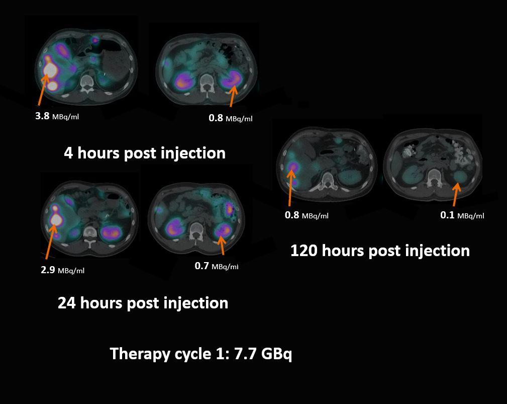
111In-Octreotide imaging with xSPECT Quant allows a more accurate assessment of neuroendocrine tumor (NET)
xSPECT Quant™ aids in neuroendocrine tumor (NET) assessment with SPECT/CT by determining changes in tumoral and non-tumoral tracer concentration in order to determine tracer retention within lesions and washout in critical organs.
Data courtesy of Royal North Shore Hospital, Sydney, Australia.
Sequential xSPECT Quant studies determine tracer retention for absorbed dose calculation
Sequential SPECT/CT with xSPECT Quant™, performed at multiple time points, was used to assess renal and tumor absorbed dose. Studies showed significant response of tumor with shrinkage ,as well as significant reduction in uptake, compared to previous study suggesting positive response to radionuclide therapy.
Data courtesy on file.
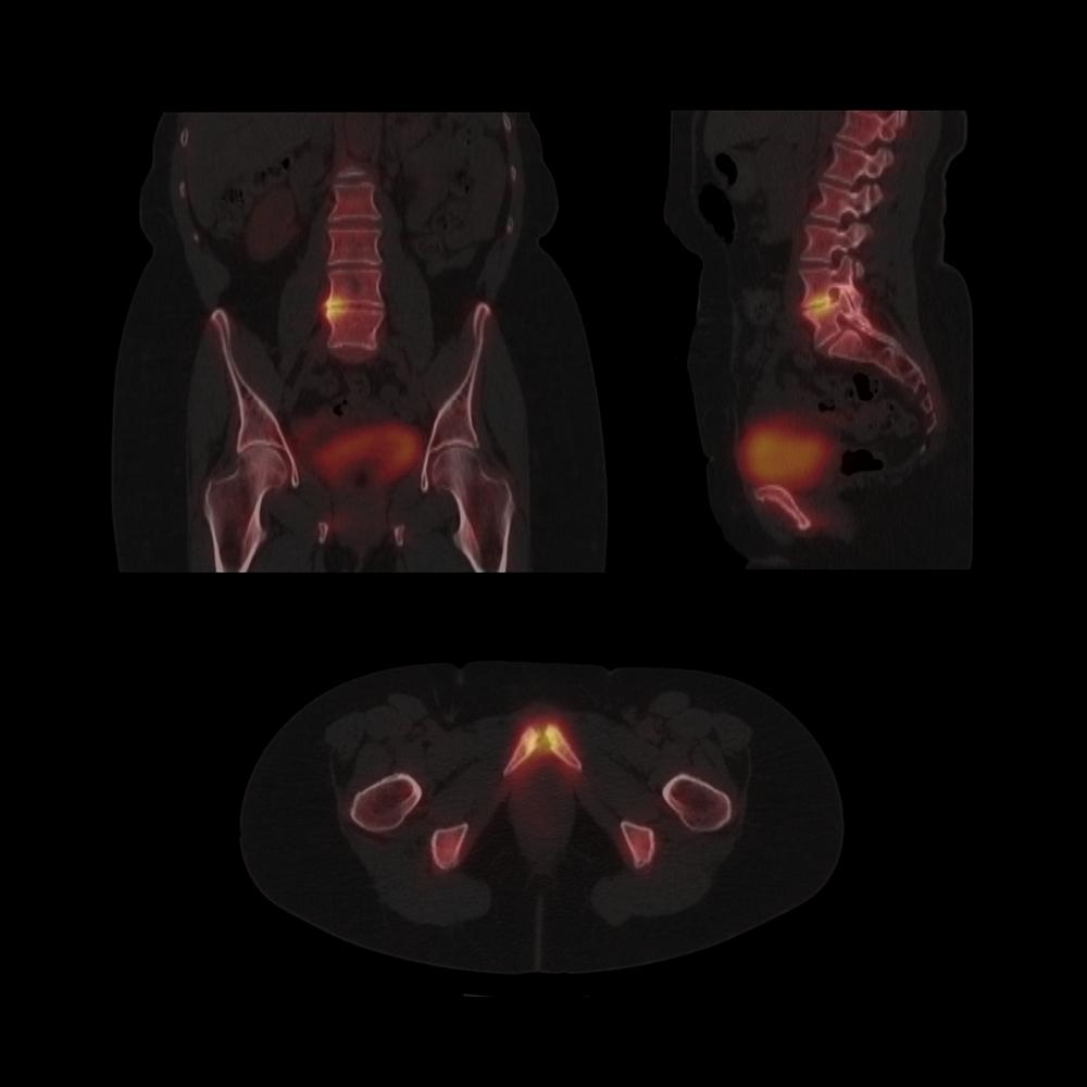
SPECT/CT aids in detection of vertebral and pelvic degenerative and inflammatory skeletal pathology
A SPECT/CT study was conducted for evaluation of skeletal pathology. SPECT/CT images confirmed increased radiographic tracer uptake. These findings are consistent with osteitis pubis and degenerative changes of the lower lumbar spine.

Data courtesy of West Virgina University Medical Center, Morgantown, West Virgina, USA.
xSPECT Bone sharply delineates right pars and laminar fracture in pediatric patient
xSPECT Bone™ shows sharp delineation of focal increased uptake corresponding to a chronic left L5 pars defect, as well as an incompletely healed fracture in the right lamina of the L5 vertebra.
Data courtesy of Groene Hart Hospital, Gouda, The Netherlands.
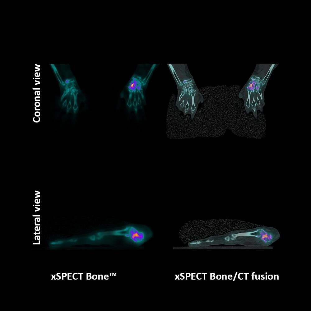
xSPECT Bone shows excellent anatomical detail of the carpal bones in patient with scaphoid fracture
xSPECT Bone™ sharply delineates hypermetabolism at the scaphoid articular surface of the scaphoid-trapezium joint, which reflects bone remodeling secondary to adjacent healed scaphoid fracture following trauma.

Data courtesy of Korea Presbyterian Medical Center, Jeonju, Jeonbuck, Republic of Korea.
xSPECT Bone delineates severity and extent of intertrochanteric hip fracture
A SPECT/CT study to assess the severity and extent of fracture and the status of right femoral head. xSPECT Bone™ sharply delineates the fracture lines, including fragmented bony margins, thereby helping visualize the displaced fragments and their relationship to adjacent soft tissue.

Data courtesy of Universitat Wurzburg, Wurzburg, Germany.
SPECT/CT helps evaluate for inducible ischemia in a patient with a history of coronary disease
A SPECT/CT myocardial perfusion scan showed no sign of significant stress-induced ischemia or transmural scars, nor inferior and posterior wall attenuation with CTAC. The normal ejection fraction (EF) was 78%.

Data courtesy on file.
SPECT/CT assists in accurate localization of neuroendocrine tumor metastases
SPECT/CT localizes multiple tracer-avid somatostatin receptor-rich hepatic metastases, thereby helping in accurate localization of lesions and differentiation of lesion from physiological uptake.

Data courtesy of Holy Cross Hospital, Fort Lauderdale, Florida, USA.
SPECT/CT localizes peri-auricular lymph sentinel node to aid in precise surgical removal
SPECT/CT imaging precisely defines a small peri-auricular sentinel lymph node due to accurate coregistration of anatomical information from the CT, with focal tracer uptake in the sentinel node seen on SPECT, allowing for precise surgical removal.

Data courtesy on file.
SPECT/CT accurately defines position of focal uptake to aid in localization of parathyroid adenoma
SPECT/CT-enabled localization of exact anatomical position of the parathyroid adenoma, which helped guide precise surgical exploration.

Data courtesy of Holy Cross Hospital, Fort Lauderdale, Florida, USA.
SPECT/CT helps characterize liver lesions with correlation of anatomical and functional information
CT shows focal hypodense liver lesions with absence of tracer uptake on SPECT. Correlation of anatomical information of liver lesions by CT and hepatocyte function by SPECT helps characterize the liver cysts.

Data courtesy of Centre Hospitalier Universitaire de Brest, Brest, France.
99mTc xSPECT Quant enables robust and reliable measurement of therapeutic effectiveness
xSPECT Quant™ across multiple time points quantifies the decrease of metabolic activity in the bone in a patient with metastases from prostate cancer. In addition, the uptake measured during anti-androgen therapy at the three time points correlated with a decline of prostate-specific antigen (PSA).

Data courtesy of University Hospital of Lausanne, Lausanne, Switzerland.
xSPECT Bone and 99mTc xSPECT Quant demonstrate details of lytic giant-cell tumor in upper fibula
xSPECT Bone™ and xSPECT Quant™ show grossly increased skeletal metabolism in bony mass (SUVmax 91.7) in the upper part of right fibula. The pattern of well-defined focal lytic areas on the CT, with heterogeneous uptake pattern on xSPECT Bone, is strongly suggestive of giant cell tumor of proximal fibula.

Data courtesy of University Hospital of Lausanne, Lausanne, Switzerland.
123I xSPECT Quant enables standardized quantification for appropriate diagnosis in equivocal motion disorders
In addition to a visual read of a SPECT/CT study, xSPECT Quant™ using 123I in 123I-Ioflupane enables standardized quantification assessment to aid in diagnosis in equivocal motion disorders.

Data courtesy of University Hospital of Lausanne, Lausanne, Switzerland.
111In-Octreotide imaging with xSPECT Quant allows a more accurate assessment of neuroendocrine tumor (NET)
xSPECT Quant™ aids in neuroendocrine tumor (NET) assessment with SPECT/CT by determining changes in tumoral and non-tumoral tracer concentration in order to determine tracer retention within lesions and washout in critical organs.

Data courtesy of Royal North Shore Hospital, Sydney, Australia.
Sequential xSPECT Quant studies determine tracer retention for absorbed dose calculation
Sequential SPECT/CT with xSPECT Quant™, performed at multiple time points, was used to assess renal and tumor absorbed dose. Studies showed significant response of tumor with shrinkage ,as well as significant reduction in uptake, compared to previous study suggesting positive response to radionuclide therapy.

Data courtesy on file.
SPECT/CT aids in detection of vertebral and pelvic degenerative and inflammatory skeletal pathology
A SPECT/CT study was conducted for evaluation of skeletal pathology. SPECT/CT images confirmed increased radiographic tracer uptake. These findings are consistent with osteitis pubis and degenerative changes of the lower lumbar spine.

Data courtesy of West Virgina University Medical Center, Morgantown, West Virgina, USA.
xSPECT Bone sharply delineates right pars and laminar fracture in pediatric patient
xSPECT Bone™ shows sharp delineation of focal increased uptake corresponding to a chronic left L5 pars defect, as well as an incompletely healed fracture in the right lamina of the L5 vertebra.

Data courtesy of Groene Hart Hospital, Gouda, The Netherlands.
xSPECT Bone shows excellent anatomical detail of the carpal bones in patient with scaphoid fracture
xSPECT Bone™ sharply delineates hypermetabolism at the scaphoid articular surface of the scaphoid-trapezium joint, which reflects bone remodeling secondary to adjacent healed scaphoid fracture following trauma.

Data courtesy of Korea Presbyterian Medical Center, Jeonju, Jeonbuck, Republic of Korea.
xSPECT Bone delineates severity and extent of intertrochanteric hip fracture
A SPECT/CT study to assess the severity and extent of fracture and the status of right femoral head. xSPECT Bone™ sharply delineates the fracture lines, including fragmented bony margins, thereby helping visualize the displaced fragments and their relationship to adjacent soft tissue.

Data courtesy of Universitat Wurzburg, Wurzburg, Germany.
SPECT/CT helps evaluate for inducible ischemia in a patient with a history of coronary disease
A SPECT/CT myocardial perfusion scan showed no sign of significant stress-induced ischemia or transmural scars, nor inferior and posterior wall attenuation with CTAC. The normal ejection fraction (EF) was 78%.


CT capability
HD detectorsAUTOFORM™ collimatorsPatient positioning monitorInnovative bed designFlash 3D iterative reconstructionCARE Dose4DAutomatic Quality Control (AQC) &
Automatic Collimator Changer (ACC)
xSPECT technology
IQ●SPECT™Technical Details
Tunnel opening
70 cm
Tunnel length
89 cm
Generator power
50 kW
Rotation time
up to 0.5 s
Tube voltage
80, 110, 130 kv
Crystal thickness
3/8” or 5/8”
Detector dimension (FOV)
53.3 x 38.7 cm
Energy range
35-588 keV
System sensitivity (LEHR at 10 cm)
202 cpm/μCi
Acquisition modes
Static, dynamic, gated, SPECT, gated SPECT,
dynamic SPECT, whole-body, whole-body SPECT,
SPECT/CT, xSPECT™Quantitative accuracy
≤ 5%2,3


