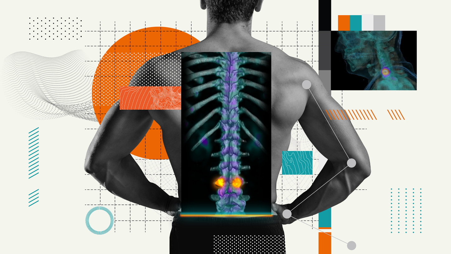
The convergence of nuclear medicine and musculoskeletal medicine has many benefits, particularly in orthopedic surgery. The capabilities of SPECT/CT technology, combined with advanced reconstruction and reading solutions, allow for accurate localization of pain origins that support diagnostic confidence and surgical accuracy.

Since 2010, Human Adams, MD, has been raising awareness of the important role nuclear medicine can play in orthopedic surgery. Adams, currently a physician in the nuclear medicine department at Groene Hart Hospital (GHZ) in Gouda, The Netherlands, became the youngest nuclear medicine physician in the country when he was just 29 years old.
After six years of practicing medicine in Gouda, Adams remains convinced of the diagnostic accuracy nuclear medicine can offer when localizing origins of pain in orthopedic patients and accurately determining the cause. He believes the hybrid imaging capability of SPECT/CT—combined with advanced reconstruction and volume-rendered visualization techniques, viewed on a dedicated reading solution—has the potential to enhance diagnostic precision, resulting in guiding either accurate orthopedic surgeries or avoiding unnecessary surgical intervention.

The clarity of nuclear medicine
In the meeting room two monitor screens light up, offering a clear overview of three different types of imaging: 2D CT images, 3D volume-rendered SPECT/CT views, and standard X-rays. As an example of the limitations of 2D images, Adams refers to a patient in recovery after hip surgery. “Osteoblastic-active areas shown in a 2D view may refer to a potential pain generator, such as the loosening of a hip prosthesis, that in reality is due to normal physiological effects of prosthesis fixation.”
Yet, as Adams begins to describe how well the combination of fused SPECT/CT images reconstructed with xSPECT Bone™ software and viewed on the syngo®.via reading solution work together, his enthusiasm shows. “This combination can increase accuracy and offer more information that is critical, for instance, in the follow-up after prosthetic loosening. Data acquired on single-modality tomography devices and viewed on conventional PACS [picture archiving and communication system] lack the pinpoint accuracy SPECT/CT images reconstructed with xSPECT Bone offer,” he explains. “For instance, with volume-rendered views of fused xSPECT Bone/CT data examined on syngo.via, we are able to define whether the osteoblastic-active area can be explained as normal physiological effects or as abnormal activity. In some cases, the cause of activity is so apparent in the images that our orthopedic surgeon is immediately convinced, and this has real added value.”

A multidisciplinary approach
In GHZ’s orthopedic department, surgeons still largely gather patient data with technology that is restricted to 2D views. Every two weeks, orthopedic surgeon Vincent Wennemers, MD, holds a multidisciplinary meeting with colleagues from his department, radiologists, and Adams to discuss orthopedic cases. Wennemers has been a surgeon at GHZ since 2016 and previously specialized in hip surgery at the Reinier de Graaf Hospital in Delft, The Netherlands.
Foot and ankle surgery, as well as hip and knee prostheses, no longer hold any secrets for him. “In general, we diagnose by means of 2D CT, X-ray, or MRI images available in the patient file via PACS. Our hospital often deals with patients who have small-joint injuries or loosened prostheses. But in order to determine whether a prosthesis has actually loosened, we consult with our colleague, Dr. Adams, and find value in the clarity his nuclear medicine images can provide.”
"In some cases, the cause of activity is so apparent in the images that our orthopedic surgeon is immediately convinced, and this has real added value."
Confidence in diagnostics
Adams reinforces, “the 2D CT, X-ray, and MRI images are of great help in orthopedic practice, but SPECT/CT images reconstructed with xSPECT Bone and viewed on syngo.via add valuable information and increase diagnostic confidence. Not all osteoblastic bone activity showcased is abnormal activity. As a result, in some cases, unnecessary surgery can be avoided,” he concludes. “My interpretation of these SPECT/CT images is that they can potentially make an orthopedic surgeon change their mind and avoid surgery. And those patients who avoid surgery can be reassured that their pain is part of the natural recovery process.”
About the author
Erika Claessens has contributed as a journalist and editor to numerous print and online publications in Belgium and the Netherlands. Her principal topics are entrepreneurial innovation and technology. She works from Antwerp, Belgium.
Excellent anatomical detail of the hands and wrist visualized with SPECT/CT
Data courtesy of Groene Hart Hospital, Gouda, The Netherlands
SPECT/CT, reconstructed with xSPECT Bone, sharply defines cortical margins and joint spaces of carpal and metacarpal bones, especially focal hyper-metabolism in the scaphoid articular surface of the scaphoid-trapezium joint, which reflects bone remodeling secondary to adjacent healed scaphoid fracture following trauma.


