AI-Rad CompanionProviding multi-modality imaging decision support

AIMed is the driving force ensuring the healthcare sector is not left behind. Their goals are to eradicate challenges, define AI enabled solutions and create an efficient workplace, with patient outcomes at its core. Their aim is to assist medical professionals to discover new ways to incorporate advances in technology to help the way they work.
Dr. Máté (Matt) Maros, neuroradiologist at Heidelberg University and user of the AI-Rad Companion Brain MR, will be one of the speakers at the virtual AIMed event “Surgery, ICU and Neurosciences” taking place on the 30-31st March. "AI-based neuroimaging: a companion for precision medicine", is the topic he will touch base on.
50% off for Siemens Healthineers customers. Please use the following discount code: AIRADQ150 (Type in the coupon code during the checkout process to receive 50% off).
The AI-Rad Companion, our family of AI-powered, cloud-based augmented workflow solutions, helps you to reduce the burden of basic repetitive tasks and may increase your diagnostic precision when interpreting medical images.
Its solutions provide automatic post-processing of imaging datasets through our AI-powered algorithms. The automation of routine workflows with repetitive tasks and high case volumes helps you to ease your daily workflow – so that you can focus on more critical issues.
Training of algorithms for AI-Rad Companion Chest CT

Training of algorithms for AI-Rad Companion Chest CT

Training of algorithms for AI-Rad Companion Chest CT
Up-to-date algorithms and scalable offerings
Our state-of-the-art algorithms will be automatically distributed to you as a user as soon as they are officially released and made available. Once the images are post-processed by the AI-Rad Companion, it supports your interpretation of the data by automatically providing results of its analysis to you for review, confirmation and possible inclusion in the final report or care pathway – to raise your precision and ensure high quality outcome in diagnostic decision-making.
All our AI-Rad Companion extensions are deployed via the teamplay digital health platform. This approach eases regular updates as well as upgrade processes and facilitates the integration of new offerings into the existing IT environment.
The AI-Rad Companion family is growing

How customers use AI-Rad Companion
Customers around the world are using AI-Rad Companion in their clinical routine. Learn how they benefit since they started using AI-Rad Companion Chest CT in their daily work.
Clinical Outcomes
AI-Rad Companion may help you to increase precision and speed up your workflow
We developed the AI-Rad Companion to help you cope with a growing workload. With its deep learning algorithms, AI-Rad Companion automatically highlights abnormalities, segments anatomies, and compares results to reference values.
Discover how AI-Rad Companion supports you as a full-time radiology expert
The AI-Rad Companion Organs RT automatically contours the organs at risk with the support of deep learning algorithms. It may reduce unwarranted variations with high-quality contours that approach the level of consensus-based contours.
Courtesy
of Leopoldina-Krankenhaus der Stadt Schweinfurt GmbH, Schweinfurt, Germany

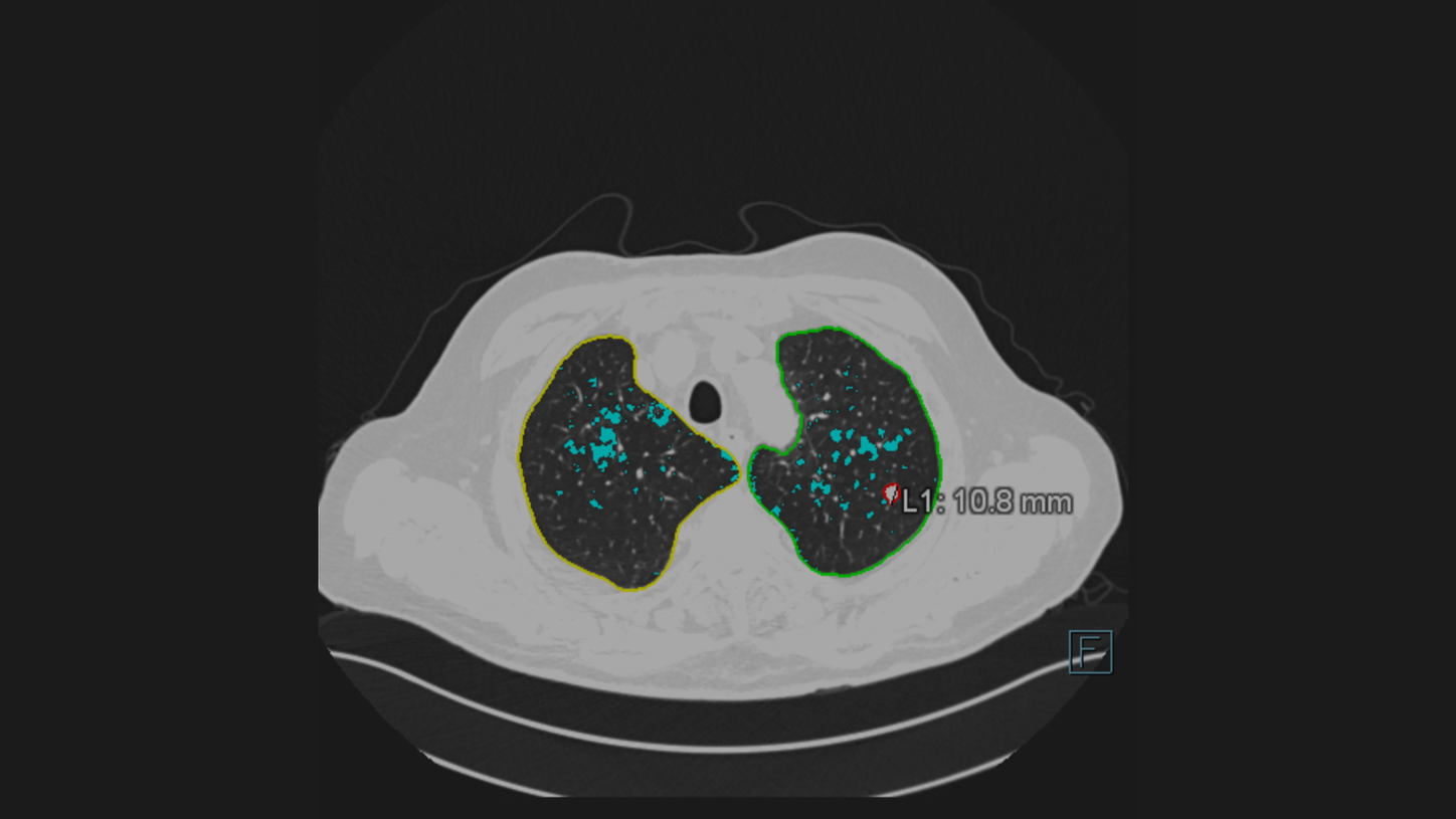
The AI-Rad Companion Chest CT detects and highlights lung nodules. Automatically after segmentation of the lung lesions, the volume, maximum 2D diameter, maximum 3D diameter and tumor burden is calculated.
Courtesy of Klinikum Nürnberg, Nuremberg, Germany

Cinematic Rendering adds clarity to the location of a detected lung nodule. The visualization makes it easy for the referring physician to get a clear picture.
Courtesy of Klinikum Nürnberg, Nuremberg, Germany

The AI-Rad Companion Chest CT recognizes automatically coronary calcium, calculates its volume and creates an enhanced clinical image including a quantification of the heart volume. This information about the cardiac condition is a valuable add-on clinical knowledge to a routine Chest CT.
Courtesy of Klinikum Nürnberg, Nuremberg, Germany

This 3D overview of the thoracic aorta has been automatically created by the AI-Rad Companion Chest CT. It includes the measurement of relevant diameters, based on medical guidelines and detected anatomical landmarks. This illustrates how the AI-Rad Companion Chest CT can support you increase the accuracy of your reporting.

The AI-Rad Companion Chest CT has automatically created this measurement of the aorta based on a non-contrast enhanced CT image.
Courtesy of Klinikum Nürnberg, Nuremberg, Germany

On this image you can see how the height of the vertebrae bodies is measured and compared. The AI-Rad Companion Chest CT automatically processes these measures and marks abnormalities with an easy to understand color-coding. Deviation are highlighted and on top you can see that the bone density is defined in Hounsfield Units.
Courtesy of Klinikum Nürnberg, Nuremberg, Germany

The new functionality Pulmonary Density3 automatically identifies and quantifies hyperdense areas of the lung using non-contrast enhanced chest CT’s. The opacity score helps to assess the percentage of affected lung tissue.

Certain parts of the pulmonary density functionality1 of AI-Rad Companion Chest CT were used in the prototype Siemens Healthineers offered to fight COVID-19, to analyze ground-glass opacities and consolidations. High opacity abnormalities were shown to correlate with lungs of COVID-19 patients.
For this sub mSv CT examination the Tin Filter technology is used.
kV: Sn130 kV, Q. ref mAs: 40 mAs, CTDIvol: 1,38 mGy DLP: 51
Eff. Dose: 0,86 mSv
Courtesy of CHR East Belgium, Verviers, Belgium

A 360° VRT overview visualizes the affected areas.
Courtesy of CHR East Belgium, Verviers, Belgium

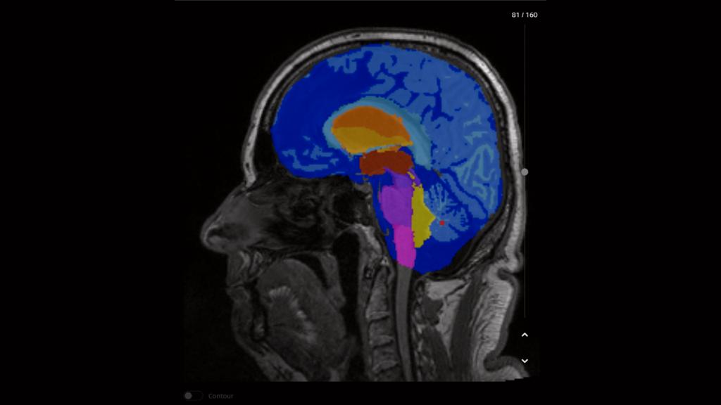
AI-Rad Companion performs an automatic segmentation of the different brain areas, including an individual volumetric analysis.
Courtesy of CHUV, Lausanne, Switzerland, Case 2aaaa1366

AI-Rad Companion compares the different volumes to a normative database and automatically generates a highlighted deviation map.
Courtesy of CHUV, Lausanne, Switzerland, Case 2aaaa1366

AI-Rad Companion automatically generates a report with all volumetric analyses listed, deviations from the normative database are marked. All results can be transferred to PACS based on the configuration.
Courtesy of Centre d'imagerie diagnostique, Lausanne, Switzerland, Case 2aaaa1366

AI-Rad Companion Prostate MR for biopsy support performs an automated segmentation as well as an automated volume estimation of the prostate. In case the PSA value is known, it calculates the PSA density.
Courtesy of Clinica Universidad de Navarra, Spain

The radiologist can manually mark and characterize lesions and other targets and add comments within the AI-Rad Companion Prostate MR for biopsy support. The solution can export segmentations and targets in a RT structured format as well as burnt in contours. These images can be exported and used by the urologist to fuse with Ultrasound images as a biopsy guidance.
Courtesy of Clinica Universidad de Navarra, Spain

PA chest X-ray of a patient showing AI-Rad Companion Chest X-ray findings of two lung lesions in the left lung and the consolidation in the right lung.
Courtesy of MVZ Prof. Dr. Uhlenbrock & Partner GmbH, Dortmund, Germany

PA chest X-ray of a patient showing an AI-Rad Companion Chest X-ray lung lesion finding in the right lung and
atelectasis in the left lung
Courtesy of MVZ Prof. Dr. Uhlenbrock & Partner GmbH, Dortmund, Germany

CT scan of the pelvis with AI-Rad Companion Organs RT generated contouring of organs at risk.
Courtesy
of University Hospital Erlangen, Erlangen, Germany

3D rendering of the CT image in the TPS showing AI-Rad Companion Organs RT generated contouring of organs at risk.
Courtesy of MVZ InnMed Oberaudorf, Germany
Rendering not generated in AI-Rad Companion Organs RT.
The AI-Rad Companion Organs RT automatically contours the organs at risk with the support of deep learning algorithms. It may reduce unwarranted variations with high-quality contours that approach the level of consensus-based contours.
Courtesy
of Leopoldina-Krankenhaus der Stadt Schweinfurt GmbH, Schweinfurt, Germany

The AI-Rad Companion Chest CT detects and highlights lung nodules. Automatically after segmentation of the lung lesions, the volume, maximum 2D diameter, maximum 3D diameter and tumor burden is calculated.
Courtesy of Klinikum Nürnberg, Nuremberg, Germany

Cinematic Rendering adds clarity to the location of a detected lung nodule. The visualization makes it easy for the referring physician to get a clear picture.
Courtesy of Klinikum Nürnberg, Nuremberg, Germany

The AI-Rad Companion Chest CT recognizes automatically coronary calcium, calculates its volume and creates an enhanced clinical image including a quantification of the heart volume. This information about the cardiac condition is a valuable add-on clinical knowledge to a routine Chest CT.
Courtesy of Klinikum Nürnberg, Nuremberg, Germany

This 3D overview of the thoracic aorta has been automatically created by the AI-Rad Companion Chest CT. It includes the measurement of relevant diameters, based on medical guidelines and detected anatomical landmarks. This illustrates how the AI-Rad Companion Chest CT can support you increase the accuracy of your reporting.

The AI-Rad Companion Chest CT has automatically created this measurement of the aorta based on a non-contrast enhanced CT image.
Courtesy of Klinikum Nürnberg, Nuremberg, Germany

On this image you can see how the height of the vertebrae bodies is measured and compared. The AI-Rad Companion Chest CT automatically processes these measures and marks abnormalities with an easy to understand color-coding. Deviation are highlighted and on top you can see that the bone density is defined in Hounsfield Units.
Courtesy of Klinikum Nürnberg, Nuremberg, Germany

The new functionality Pulmonary Density3 automatically identifies and quantifies hyperdense areas of the lung using non-contrast enhanced chest CT’s. The opacity score helps to assess the percentage of affected lung tissue.

Certain parts of the pulmonary density functionality1 of AI-Rad Companion Chest CT were used in the prototype Siemens Healthineers offered to fight COVID-19, to analyze ground-glass opacities and consolidations. High opacity abnormalities were shown to correlate with lungs of COVID-19 patients.
For this sub mSv CT examination the Tin Filter technology is used.
kV: Sn130 kV, Q. ref mAs: 40 mAs, CTDIvol: 1,38 mGy DLP: 51
Eff. Dose: 0,86 mSv
Courtesy of CHR East Belgium, Verviers, Belgium

A 360° VRT overview visualizes the affected areas.
Courtesy of CHR East Belgium, Verviers, Belgium

AI-Rad Companion performs an automatic segmentation of the different brain areas, including an individual volumetric analysis.
Courtesy of CHUV, Lausanne, Switzerland, Case 2aaaa1366

AI-Rad Companion compares the different volumes to a normative database and automatically generates a highlighted deviation map.
Courtesy of CHUV, Lausanne, Switzerland, Case 2aaaa1366

AI-Rad Companion automatically generates a report with all volumetric analyses listed, deviations from the normative database are marked. All results can be transferred to PACS based on the configuration.
Courtesy of Centre d'imagerie diagnostique, Lausanne, Switzerland, Case 2aaaa1366

AI-Rad Companion Prostate MR for biopsy support performs an automated segmentation as well as an automated volume estimation of the prostate. In case the PSA value is known, it calculates the PSA density.
Courtesy of Clinica Universidad de Navarra, Spain

The radiologist can manually mark and characterize lesions and other targets and add comments within the AI-Rad Companion Prostate MR for biopsy support. The solution can export segmentations and targets in a RT structured format as well as burnt in contours. These images can be exported and used by the urologist to fuse with Ultrasound images as a biopsy guidance.
Courtesy of Clinica Universidad de Navarra, Spain

PA chest X-ray of a patient showing AI-Rad Companion Chest X-ray findings of two lung lesions in the left lung and the consolidation in the right lung.
Courtesy of MVZ Prof. Dr. Uhlenbrock & Partner GmbH, Dortmund, Germany

PA chest X-ray of a patient showing an AI-Rad Companion Chest X-ray lung lesion finding in the right lung and
atelectasis in the left lung
Courtesy of MVZ Prof. Dr. Uhlenbrock & Partner GmbH, Dortmund, Germany

CT scan of the pelvis with AI-Rad Companion Organs RT generated contouring of organs at risk.
Courtesy
of University Hospital Erlangen, Erlangen, Germany

3D rendering of the CT image in the TPS showing AI-Rad Companion Organs RT generated contouring of organs at risk.
Courtesy of MVZ InnMed Oberaudorf, Germany
Rendering not generated in AI-Rad Companion Organs RT.
The AI-Rad Companion Organs RT automatically contours the organs at risk with the support of deep learning algorithms. It may reduce unwarranted variations with high-quality contours that approach the level of consensus-based contours.
Courtesy
of Leopoldina-Krankenhaus der Stadt Schweinfurt GmbH, Schweinfurt, Germany


Integration
Smooth integration of artificial intelligence into the radiology environment
Drive productivity with seamless integration in the reading and reporting workflow includingautomated measurements and DICOM structured reports ̶ while every workflow step remains under control to enable evidence based decisions.

COVID-19

The entire global population has been impacted, in one way or another, by the COVID-19 virus. The AI-Rad Companion, our Siemens Healthineers AI driven solution, offers our customers the opportunity to have access to different postprocessing software for the analysis of lung examinations.
AI-Rad Companion Research Chest X-ray Pneumonia Analysis4
This prototype is designed to automatically identify and quantify airspace opacities of the lung, enabling simple to use analysis of chest X-rays for research purposes only (not for clinical use). AI-Rad Companion Research Chest X-ray Pneumonia Analysis will perform automated lung opacity analysis on Chest X-ray (AP or PA viewing direction), highlighting identified regions. Other radiographical findings, such as pneumothorax, pulmonary lesions, atelectasis, consolidation and pleural effusion are included as well.
AI-Rad Companion Chest X-ray2
Chest imaging, X-ray in particular, plays an important role in patient management during the COVID-19 pandemic5. Patient management and clinical decisions depend on clinical outcomes and imaging reports. The new member of the AI-Rad Companion family, the AI-Rad Companion Chest X-ray, automatically processes upright chest X-ray images (PA direction). Next to pneumothorax, pleural effusion and nodule detection, the AI-Rad Companion Chest X-ray is able to indicate consolidations and atelectasis. The latter may be signs of pneumonia caused by the COVID-19 virus.
AI-Rad Companion Brain MR, Chest CT, Prostate MR and Organs RT are not yet commercially available in all countries and their future availability cannot be ensured.
AI-Rad Companion Chest X-ray is currently under development; it is not for sale in the United States and other countries. CE mark is available.
The pulmonary density functionality is not commercially available in all countries, and its future availability cannot be ensured. As soon as this functionality is available and cleared for your country it will become entitled to your institution.
The Pulmonary Density feature is new in VA12A without FDA Clearance. According to FDA policy “Enforcement Policy for Imaging Systems During the Coronavirus Disease 2019 (COVID-19) Public Health Emergency” issued in April 2020, the manufacturer is allowed to market this feature without FDA-clearance. This policy is intended to remain in effect only for the duration of the public health emergency related to COVID-19 declared by the HHS, including any renewals made by the HHS Secretary in accordance with section 319(a)(2) of the Public Health Services Act (42 U.S.C. 247d(a)(2)). Pulmonary Density results are not indicated for the diagnosis of COVID-19. Only in vitro diagnostic testing is currently the definitive method to diagnose COVID-19.
For research purpose only. Not for clinical use. This prototype is still under development and not yet commercially available. Its future availability cannot be ensured.
https://pubs.rsna.org/doi/10.1148/radiol.2020201365
Attenzione: Siemens Healthineers è consapevole che misuratori di glucosio non invasivi e ad uso domestico sono offerti in vendita su numerosi siti e social network. Questi prodotti non sono sviluppati né prodotti né distribuiti da Siemens Healthineers. Chi promuove e distribuisce tali prodotti, che non provengono, né sono riferibili o autorizzati da Siemens Healthineers, sta utilizzando impropriamente ed illegalmente il brand dell’azienda. Se vi siete imbattuti in un annuncio o in una pagina web di questo tipo, segnalateci i dettagli via e-mail a: healthcare.it.communications.rc-it.team@siemens-healthineers.com
Avvertenza: Accesso al sito siemens-healthineers.com/it
Le informazioni contenute in questo sito sono destinate in via esclusiva agli operatori professionali della sanità in conformità all’art. 21 del D.Lgs. 24 febbraio 1997, n. 46 s.m.i e alle Linee Guida del Ministero della Salute del 17 febbraio 2010 e successivo aggiornamento del 18 marzo 2013.
Per proseguire cliccare ’OK’ per uscire cliccare ’Annulla’.
Il sistema ricorderà la vostra scelta.

