Symbia IntevoSPECT/CT that improves your image
Discover expanded clinical reach and a new standard of care. Symbia Intevo™ combines the improved image quality and localization of CT with proven SPECT capabilities, resulting in hybrid SPECT/CT technology that will distinguish your facility.

Benefits

Symbia Intevo offers high SPECT sensitivity1 and reconstructed resolution, along with high-performance CT, standard on every system. The result: confidence in knowing that every exam is performed with highest image quality.1

Symbia Intevo SPECT/CT imaging provides a more complete picture of your patient's condition in a single examination, allowing you to quickly make decisions that result in successful treatment strategies and a more satisfying experience.

Expand the scope of your imaging services with Symbia Intevo's advanced SPECT and CT technologies—and further distinguish your facility among referring physicians, patients, and the medical community through a reputation for providing quick, meaningful results.
Symbia Intevo offers high SPECT sensitivity1 and reconstructed resolution, along with high-performance CT, standard on every system. The result: confidence in knowing that every exam is performed with highest image quality.1
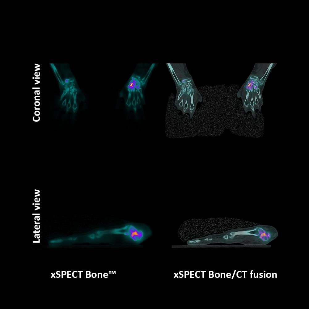
Evidence
Data courtesy on file.
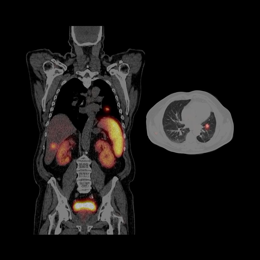
SPECT/CT assists in accurate localization of neuroendocrine tumor metastases
SPECT/CT localizes multiple tracer-avid somatostatin receptor-rich hepatic metastases, thereby helping in accurate localization of lesions and differentiation of lesion from physiological uptake.


Technical Details
Tunnel opening
70 cm
Tunnel length
89 cm
Generator power
50 kW
Rotation time
up to 0.5 s
Tube voltage
80, 110, 130 kv
Crystal thickness
3/8” or 5/8”
Detector dimension (FOV)
53.3 x 38.7 cm
Energy range
35-588 keV
System sensitivity (LEHR at 10 cm)
202 cpm/μCi
Acquisition modes
Static, dynamic, gated, SPECT, gated SPECT,
dynamic SPECT, whole-body, whole-body SPECT,
SPECT/CT, xSPECT™Quantitative accuracy
≤ 5%2,3


