Stroke Imaging
When stroke strikes, be ahead of your time.
As your partner in stroke management, we keep you ahead of your time. Our advanced technologies let you speed up stroke care and transform care delivery along the entire pathway – from stroke screening and pre-hospital diagnosis and care to in-hospital diagnosis and treatment. Explore our offerings for stroke care!
The clinical stroke pathway
You rely on excellent diagnostic imaging to confirm ischemic stroke, detect the location of the vessel occlusion and evaluate the brain damage and penumbra. CT and MR imaging are considered the gold standard by international stroke guidelines. But when every second counts, having the right software and technology for treatment of acute stroke patients matters.
1/4

4. Treatment
Connecting more people to advanced care – image-guided endovascular robotics
With image-guided, robotic-assisted therapy, we are transforming the way endovascular care is delivered. Together, advanced imaging and robotics with assisted automation enable physicians to treat patients more precisely – With our vision to reach more people and expedite time to treatment, we’ll bring essential therapy to heart attack and stroke patients where they are, when they need it.
- Telerobotics will expand access to care
- Future technology integration will improve workflow and patient outcomes
- Specialized capabilities make treatment safer and more precise
- Innovations create a safer environment

1. CT Imaging
Recon&GO is a ready-to-read technology. With AI recognizing patient landmarks and anatomies, Recon&GO automates postprocessing tasks and becomes part of the standard reconstruction tasks.
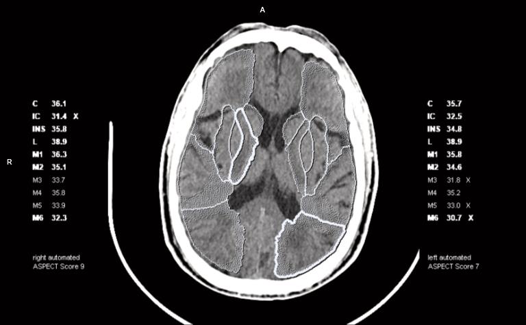
For stroke patients, Recon&GO Inline ASPECTS automatically calculates the ASPECT score of a non-contrast CT head scan.
The affected brain regions are automatically highlighted as an overlay on the CT image based on a quantitative topographic comparison of the left and right hemisphere. Automatic segmentation into 10 cerebral regions based on imaging data of patients who presented with symptoms of acute stroke with proven vascular occlusion.
The images and results are automatically calculated in the background and can be directly sent to PACS without any user interaction. This makes Inline ASPECTS routine-ready by providing consistent results independent of the user and always available especially in urgent situations when time is a scarce resource.
When a perfusion scan is performed, syngo.CT Neuro Perfusion provides an automated workflow in which the perfusion maps, tissue-at-risk (penumbra) and potential recuperation ratio (PRR) are calculated. As for Recon&GO Inline ASPECTS, these results can be directly sent to PACS without any user interaction.

2. MR Imaging
Speed is key since “Brain is Time”, so fast patient preparation and fast scanning in MR imaging is important.
With AutoAlign Head LS we can speed up the patient preparation
Automated positioning and alignment of slice groups to the anatomy, relying on multiple anatomical landmarks. Provides fast, easy, and reproducible patient scanning and facilitates the reading by consistently delivering high image quality with a standardized slice orientation, both for follow-ups and across patients. AutoAlign Head LS computes the central positioning for many routine brain structures such as AC-PC, midbrain and temporal lobes. The inner ear, the orbits and the optic nerve are also standard positioning orientations with the AutoAlign Head LS. It delivers robust and consistent results independently of patient age, head position, disease or existing lesions.
AutoAlign Head is part of myExam Brain Assist.

With the user defined perfusion parameters and threshold values, a 3D evaluation of brain tissue for the mismatch between the infarct core (red) and the penumbra (yellow) can be performed demonstrating the full extent of ischemic damage.
3. Diagnosis and IT solutions
Our comprehensive syngo.CT portfolio for stroke management enables fast and reproducible results without manual user interaction to support further triage and treatment pathways.
The syngo.CT Neuro Perfusion software package allows to evaluate areas of brain perfusion. The software processes images or volumes that were reconstructed from continuously acquired CT data after the injection of contrast media. Perfusion parameters such as Cerebral blood flow (CBF), Cerebral blood volume (CBV), Local bolus, Mean transit time (MTT), Transit time to the center of the IRF (Tmax), Flow extraction product, Temporal MIP & Average and Baseline Volume can be calculated.
The software also allows the calculation of mirrored regions or volumes of interest and the visual inspection of time attenuation curves. The software can be used to visualize the apparent blood perfusion and the parameter mismatch in brain tissue affected by acute stroke. It can also visualize blood brain barrier disturbances by modeling extravascular leakage of blood into the interstitial space. This additional capability may improve the differential diagnosis of brain tumors and be helpful in therapy monitoring.
Auto Stroke, in combination with Siemens Healthineers’ Rapid Results technology can auto-archive the automatically generated results (e.g., perfusion maps, core and penumbra volumes, etc.) to PACS without user interaction.

4. Treatment
Connecting more people to advanced care – image-guided endovascular robotics
With image-guided, robotic-assisted therapy, we are transforming the way endovascular care is delivered. Together, advanced imaging and robotics with assisted automation enable physicians to treat patients more precisely – With our vision to reach more people and expedite time to treatment, we’ll bring essential therapy to heart attack and stroke patients where they are, when they need it.
- Telerobotics will expand access to care
- Future technology integration will improve workflow and patient outcomes
- Specialized capabilities make treatment safer and more precise
- Innovations create a safer environment

1. CT Imaging
Recon&GO is a ready-to-read technology. With AI recognizing patient landmarks and anatomies, Recon&GO automates postprocessing tasks and becomes part of the standard reconstruction tasks.
For stroke patients, Recon&GO Inline ASPECTS automatically calculates the ASPECT score of a non-contrast CT head scan.
The affected brain regions are automatically highlighted as an overlay on the CT image based on a quantitative topographic comparison of the left and right hemisphere. Automatic segmentation into 10 cerebral regions based on imaging data of patients who presented with symptoms of acute stroke with proven vascular occlusion.
The images and results are automatically calculated in the background and can be directly sent to PACS without any user interaction. This makes Inline ASPECTS routine-ready by providing consistent results independent of the user and always available especially in urgent situations when time is a scarce resource.
When a perfusion scan is performed, syngo.CT Neuro Perfusion provides an automated workflow in which the perfusion maps, tissue-at-risk (penumbra) and potential recuperation ratio (PRR) are calculated. As for Recon&GO Inline ASPECTS, these results can be directly sent to PACS without any user interaction.

2. MR Imaging
Speed is key since “Brain is Time”, so fast patient preparation and fast scanning in MR imaging is important.
With AutoAlign Head LS we can speed up the patient preparation
Automated positioning and alignment of slice groups to the anatomy, relying on multiple anatomical landmarks. Provides fast, easy, and reproducible patient scanning and facilitates the reading by consistently delivering high image quality with a standardized slice orientation, both for follow-ups and across patients. AutoAlign Head LS computes the central positioning for many routine brain structures such as AC-PC, midbrain and temporal lobes. The inner ear, the orbits and the optic nerve are also standard positioning orientations with the AutoAlign Head LS. It delivers robust and consistent results independently of patient age, head position, disease or existing lesions.
AutoAlign Head is part of myExam Brain Assist.

With the user defined perfusion parameters and threshold values, a 3D evaluation of brain tissue for the mismatch between the infarct core (red) and the penumbra (yellow) can be performed demonstrating the full extent of ischemic damage.
3. Diagnosis and IT solutions
Our comprehensive syngo.CT portfolio for stroke management enables fast and reproducible results without manual user interaction to support further triage and treatment pathways.
The syngo.CT Neuro Perfusion software package allows to evaluate areas of brain perfusion. The software processes images or volumes that were reconstructed from continuously acquired CT data after the injection of contrast media. Perfusion parameters such as Cerebral blood flow (CBF), Cerebral blood volume (CBV), Local bolus, Mean transit time (MTT), Transit time to the center of the IRF (Tmax), Flow extraction product, Temporal MIP & Average and Baseline Volume can be calculated.
The software also allows the calculation of mirrored regions or volumes of interest and the visual inspection of time attenuation curves. The software can be used to visualize the apparent blood perfusion and the parameter mismatch in brain tissue affected by acute stroke. It can also visualize blood brain barrier disturbances by modeling extravascular leakage of blood into the interstitial space. This additional capability may improve the differential diagnosis of brain tumors and be helpful in therapy monitoring.
Auto Stroke, in combination with Siemens Healthineers’ Rapid Results technology can auto-archive the automatically generated results (e.g., perfusion maps, core and penumbra volumes, etc.) to PACS without user interaction.

4. Treatment
Connecting more people to advanced care – image-guided endovascular robotics
With image-guided, robotic-assisted therapy, we are transforming the way endovascular care is delivered. Together, advanced imaging and robotics with assisted automation enable physicians to treat patients more precisely – With our vision to reach more people and expedite time to treatment, we’ll bring essential therapy to heart attack and stroke patients where they are, when they need it.
- Telerobotics will expand access to care
- Future technology integration will improve workflow and patient outcomes
- Specialized capabilities make treatment safer and more precise
- Innovations create a safer environment


1/4
Future vision for stroke management
We at Siemens Healthineers are committed to helping healthcare providers globally to succeed in today’s dynamic environment. We are inspired to transform the way things are done – because we want what is best for our people, our customers, and ultimately the health of mankind.
Data integration, smart intelligent systems will be necessary to evolve the quality of treatment delivered to the patients.
Get a small taste of what future digitalization and intelligence can mean for Paula, a middle-aged woman with an elevated risk of being hit by a stroke.
Digitalizing healthcare is the key enabler for expanding precision medicine transforming care delivery, and improving patient experience. Ultimately, digitalizing healthcare enables providers to achieve better outcomes at lower costs. To make this possible, four steps are critical:
- Manage data as a strategic asset, e.g. with the support of technology like the digital twin
- Empower data-driven decisions
- Connect care teams and patients
- Build a learning health system, e.g. using AI technology
Useful links

Artis Interventional Angiography Systems


Magnetic Resonance Imaging

syngo.via
Useful documents
CT Neuro Perfusion in ischemic stroke management
Stroke treatment in the fast lane
I would like to know more on Stroke Imaging
See the other topics here
Vond u deze informatie nuttig?
Bedankt.
Wilt u ons uw feedback geven?
125 / 125
Deze pagina is uitsluitend bedoeld voor professionals uit de gezondheidszorgsector. Gelieve te bevestigen dat u een gezondheidszorgprofessional bent door op OK te klikken.
