MR imaging in sports medicineImaging excellence for all athletes: from daily fitness to peak performance
With more people leading active lifestyles, the need for MR imaging in sports medicine is growing. Our MRI technology offers valuable insights into athlete injuries, providing detailed, radiation-free images of soft tissues, joints, and bones. Mild traumatic brain injuries and cardiovascular diseases can be evaluated using MRI.
Highlights
High-speed sports often lead to sprains, fractures, and concussions, while repetitive movements frequently cause upper body injuries. MRI is crucial for diagnosing soft tissue damage and guiding treatment after concussions. For serious athletes, tools like cardiac MRI might help mitigate life-threatening cardiovascular events.

Orthopedic imaging
Provides detailed images of soft tissues, bones, and joints for injury assessment and treatment planning. It can help predict return to play of athletes.

Neuro imaging
Helps evaluating potential soft tissue injuries after mild traumatic brain injuries such as concussions or contusions.

Cardiac imaging
In competitive athletes the incidence of Sudden Cardiac Death is high. Cardiovascular MRI (CMR) is a powerful screening/ risk stratification tool.
Tailored imaging for all athletes
AI integration and automated workflows ensure fast results, while quantitative assessments enable confident treatment decisions and effective recovery monitoring.

Pioneering Innovation
Early detection of cartilage changes with Chrondal Quant.
Clinical solutions

BioMatrix Beat Sensor2
BioMatrix Beat Sensor enables users to perform CMR exams without ECG. This break-through technology uses the heart's own pulsile motion to trigger the exam. The result is faster CMR exams and improved patient experience.

Dedicated coils for dedicated needs
Our dedicated MSK coils are ergonomically designed and enable high-resolution scanning.
From our 18-channel transmit/receive knee coil with its flared opening to the Foot/Ankle 16 and its boot-like design our coils enable fast patient positioning.
Shoulder Shape 16 has flexible wings to shape around the shoulder anatomy and Hand/Wrist 16 is adapted to the shape of the hand and wrist for easy and comfortable patient positioning and high- resolution imaging.

Simplify exams of large joints with myExam Large Joint Assist
myExam Large Joint Assist simplifies the examination of all large joints (knee, shoulder, hip) and minimizes potential sources of planning errors with AutoAlignand AutoCoverage. Expand your diagnostic confidence with ready-to-use optimized protocols for artifact reduction or motion robust imaging with single-click changes to a scan strategy.
Your benefits at a glance:
- Consistent and efficient exam planning powered by artificial intelligence
- At any time, reproducible and high- quality image results
- Ready-to-use optimized protocols for artifact reduction or motion robust imaging

myExam Assist for Brain and Spine
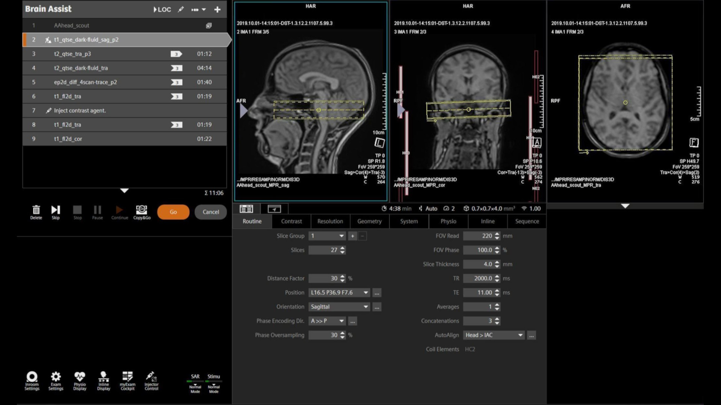
As part of myExam Companion, myExam Assist Brain makes general brain exams easier, more reproducible, and more efficient. myExam Assist Spine optimizes cervical, thoracic, and lumbar spine imaging for a wide range of patients and conditions. AutoAlign automizes slice planning along the whole CNS, automated vertebrae labeling and automatic detection of the spine geometry reduce workflow steps to streamline the exams.
Your benefits:
- Faster and more efficient patient scheduling, consistent results for follow-up exams
- Reduce wasteful workflow steps to be more patient focused
- Strategies and decision points provide clear guidance during the exam

Courtesy of SAHMRI, Adelaide, Australia | 2aaaa2892
Wave-CAIPI SWI
The perfect CAIPIRINHA: Wave-CAIPI SWI allows exploiting coil sensitivity variations in all three dimensions, thereby achieving remarkable parallel imaging acceleration factors. Obtain SWI data of the whole brain in substantially less acquisition time than before, outrun potential motion artifacts and keep patient table times at bay.
Your benefits:
- Higher acceleration factors by optimal usage of three-dimensional coil sensitivity variations
- Whole brain SWI in less than two minutes
- Most beneficial in combination with BioMatrix Head/Neck 64 channel coil with CoilShim for optimal SNR and field homogeneity
AutoMate Cardiac1
AutoMate Cardiac enhances cardiovascular MR imaging by using AI-based automated workflows to set up scan parameters, ensuring robust image quality regardless of operator expertise.
myExam Cardiac Assist
myExam Cardiac Assist is a unique offering from Siemens Healthineers which enables comprehensive Cardiac MR examinations in under 30 minutes.

BioMatrix Beat Sensor2
BioMatrix Beat Sensor enables users to perform CMR exams without ECG. This break-through technology uses the heart's own pulsile motion to trigger the exam. The result is faster CMR exams and improved patient experience.

Dedicated coils for dedicated needs
Our dedicated MSK coils are ergonomically designed and enable high-resolution scanning.
From our 18-channel transmit/receive knee coil with its flared opening to the Foot/Ankle 16 and its boot-like design our coils enable fast patient positioning.
Shoulder Shape 16 has flexible wings to shape around the shoulder anatomy and Hand/Wrist 16 is adapted to the shape of the hand and wrist for easy and comfortable patient positioning and high- resolution imaging.

Simplify exams of large joints with myExam Large Joint Assist
myExam Large Joint Assist simplifies the examination of all large joints (knee, shoulder, hip) and minimizes potential sources of planning errors with AutoAlignand AutoCoverage. Expand your diagnostic confidence with ready-to-use optimized protocols for artifact reduction or motion robust imaging with single-click changes to a scan strategy.
Your benefits at a glance:
- Consistent and efficient exam planning powered by artificial intelligence
- At any time, reproducible and high- quality image results
- Ready-to-use optimized protocols for artifact reduction or motion robust imaging

myExam Assist for Brain and Spine
As part of myExam Companion, myExam Assist Brain makes general brain exams easier, more reproducible, and more efficient. myExam Assist Spine optimizes cervical, thoracic, and lumbar spine imaging for a wide range of patients and conditions. AutoAlign automizes slice planning along the whole CNS, automated vertebrae labeling and automatic detection of the spine geometry reduce workflow steps to streamline the exams.
Your benefits:
- Faster and more efficient patient scheduling, consistent results for follow-up exams
- Reduce wasteful workflow steps to be more patient focused
- Strategies and decision points provide clear guidance during the exam

Courtesy of SAHMRI, Adelaide, Australia | 2aaaa2892
Wave-CAIPI SWI
The perfect CAIPIRINHA: Wave-CAIPI SWI allows exploiting coil sensitivity variations in all three dimensions, thereby achieving remarkable parallel imaging acceleration factors. Obtain SWI data of the whole brain in substantially less acquisition time than before, outrun potential motion artifacts and keep patient table times at bay.
Your benefits:
- Higher acceleration factors by optimal usage of three-dimensional coil sensitivity variations
- Whole brain SWI in less than two minutes
- Most beneficial in combination with BioMatrix Head/Neck 64 channel coil with CoilShim for optimal SNR and field homogeneity
AutoMate Cardiac1
AutoMate Cardiac enhances cardiovascular MR imaging by using AI-based automated workflows to set up scan parameters, ensuring robust image quality regardless of operator expertise.
myExam Cardiac Assist
myExam Cardiac Assist is a unique offering from Siemens Healthineers which enables comprehensive Cardiac MR examinations in under 30 minutes.

BioMatrix Beat Sensor2
BioMatrix Beat Sensor enables users to perform CMR exams without ECG. This break-through technology uses the heart's own pulsile motion to trigger the exam. The result is faster CMR exams and improved patient experience.


Clinical cases

Courtesy: St. Georg Hospital, Leipzig, Germany. Study-ID: 1aaaa2598
PSIR HeartFreeze4
PSIR HeartFreeze for LGE scar assessment in free-breathing
Image aquired with MAGNETOM Altea (1.5T)
Study-ID: 2aaaa2947
Hand imaging
Finger fracture scanned with Deep Resolve in Hand/Wrist 16.
Image acquired with MAGNETOM Vida (3T).
Courtesy Diagnostikzentrum Graz; Study-ID: 2aaaa2399
Knee imaging
Patella luxation and bone bruise with TX/RX knee 18 coil , Deep Resolve and Simultaneous Multi- Slice.
Images acquired with MAGNETOM Vida (3T).
Study-ID: 1aaaa4709
Shoulder imaging
Athlete post surgical supraspinatus ruptur examined with Deep Resolve and Shoulder Shape 16.
Image acquired with MAGNETOM Altea (1.5T).
Study-ID: 3aaaa1105
Hip imaging
Hip Arthrography showing a labral ruptur scanned with 3D isotropic Compressed Sensing SPACE.
By accelerating imaging with Compressed Sensing 3D images with sub-millimeter resolution for all relevant contrasts can be acquired in less than 5 minutes per scan.
Image acquired with MAGNETOM Vida (3T).
Courtesy Diagnostikzentrum Graz; Study-ID: 2aaaa2075
Spine imaging
Protusion of the l-spine using MyExam Spine Assist with spine labeling.
Image acquired with MAGNETOM Vida (3T).
Study-ID: 2aaaa2399
Ankle imaging
A talofibular-ligament rupture scanned with boot –like designed Foot/Ankle 16 and Deep Resolve.
Image acquired with MAGNETOM Vida Fit (3T).
Courtesy of Benson Radiology, Adelaide, Australia; Study-ID: 2aaaa2951
Deep Resolve for SPACE is currently under development and not commercially available in the US. Its future availability cannot be ensured
Nerve imaging
Brachial plexus with Planned Deep Resolve for SPACE.
Image acquired on MAGNETOM Vida (3T).

Study-ID: 1aaaa2587
Brain imaging
Image acquired on MAGNETOM Altea (1.5T).

Case courtesy: J. Dehem, Jan Yperman Ziekenhuis, Ypers, Belgium, Study ID 1aaaa3348
Cardiac imaging
Late gadolinium enhancement (LGE) for tissue characterization.
This was a 46 year old active tennis player with suddent cardiac arrest on the tennis court; CPR, cath lab and now CMR as follow up.
Siemens Healthineers. Study-ID: 2aaaa1877
BioMatrix Beat Sensor2
Comparison: Cardiac triggering with ECG versus cardiac triggering using Beat Sensor.
Image aquired with MAGNETOM Sola (1.5T)

Courtesy: St. Georg Hospital, Leipzig, Germany. Study-ID: 1aaaa2598
PSIR HeartFreeze4
PSIR HeartFreeze for LGE scar assessment in free-breathing
Image aquired with MAGNETOM Altea (1.5T)
Study-ID: 2aaaa2947
Hand imaging
Finger fracture scanned with Deep Resolve in Hand/Wrist 16.
Image acquired with MAGNETOM Vida (3T).
Courtesy Diagnostikzentrum Graz; Study-ID: 2aaaa2399
Knee imaging
Patella luxation and bone bruise with TX/RX knee 18 coil , Deep Resolve and Simultaneous Multi- Slice.
Images acquired with MAGNETOM Vida (3T).
Study-ID: 1aaaa4709
Shoulder imaging
Athlete post surgical supraspinatus ruptur examined with Deep Resolve and Shoulder Shape 16.
Image acquired with MAGNETOM Altea (1.5T).
Study-ID: 3aaaa1105
Hip imaging
Hip Arthrography showing a labral ruptur scanned with 3D isotropic Compressed Sensing SPACE.
By accelerating imaging with Compressed Sensing 3D images with sub-millimeter resolution for all relevant contrasts can be acquired in less than 5 minutes per scan.
Image acquired with MAGNETOM Vida (3T).
Courtesy Diagnostikzentrum Graz; Study-ID: 2aaaa2075
Spine imaging
Protusion of the l-spine using MyExam Spine Assist with spine labeling.
Image acquired with MAGNETOM Vida (3T).
Study-ID: 2aaaa2399
Ankle imaging
A talofibular-ligament rupture scanned with boot –like designed Foot/Ankle 16 and Deep Resolve.
Image acquired with MAGNETOM Vida Fit (3T).
Courtesy of Benson Radiology, Adelaide, Australia; Study-ID: 2aaaa2951
Deep Resolve for SPACE is currently under development and not commercially available in the US. Its future availability cannot be ensured
Nerve imaging
Brachial plexus with Planned Deep Resolve for SPACE.
Image acquired on MAGNETOM Vida (3T).

Study-ID: 1aaaa2587
Brain imaging
Image acquired on MAGNETOM Altea (1.5T).

Case courtesy: J. Dehem, Jan Yperman Ziekenhuis, Ypers, Belgium, Study ID 1aaaa3348
Cardiac imaging
Late gadolinium enhancement (LGE) for tissue characterization.
This was a 46 year old active tennis player with suddent cardiac arrest on the tennis court; CPR, cath lab and now CMR as follow up.
Siemens Healthineers. Study-ID: 2aaaa1877
BioMatrix Beat Sensor2
Comparison: Cardiac triggering with ECG versus cardiac triggering using Beat Sensor.
Image aquired with MAGNETOM Sola (1.5T)

Courtesy: St. Georg Hospital, Leipzig, Germany. Study-ID: 1aaaa2598
PSIR HeartFreeze4
PSIR HeartFreeze for LGE scar assessment in free-breathing
Image aquired with MAGNETOM Altea (1.5T)


