- Siemens Healthineers Polska
- Medical Imaging
- Obrazowanie molekularne
- PET/CT
- Digital LSO-based PET detector

Set the standard in PET/CT
Optiso HD LSO-based detectors advance PET/CT imaging

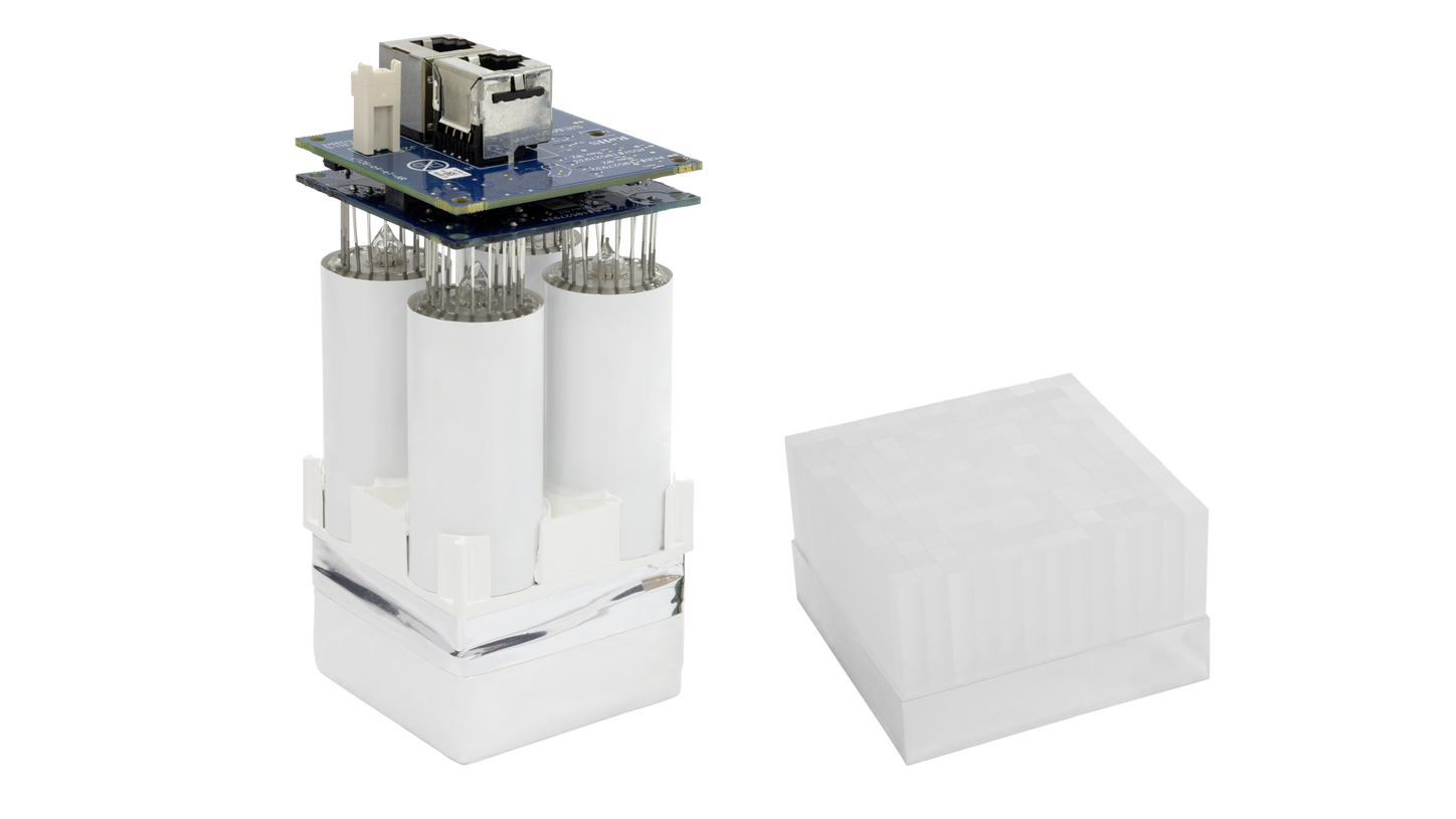
Optiso HD digital detector
For True time of flight and better image quality
We are the sole PET manufacturer to own the entire manufacturing process for the detector. This allows us to closely monitor process quality control and gives us the flexibility to explore different detector designs to reach an optimal detector solution.
What sets us apart:
- Crystals are grown, cut, and individually selected in-house
- Self-manufactured ensures conformance to precise specifications and allows for a deep understanding of optimal detector design
- Higher light output and faster scintillation,1 enable true time of flight (TOF) and better image quality
Setting the standard in PET/CT with digital LSO-based detectors
Biograph™ mCT and Biograph Horizon digital detectors offer PET/CT imaging with high, 78-mm3 volumetric resolution2 due to the small, 4 x 4-mm lutetium oxyorthosilicate (LSO) crystal elements and the ability to offer true TOF for performance and clinical advantages.

4.0-mm LSO crystals
For improved and more precise imaging
In the early 2000s, we took the initiative of replacing all of our BGO PET/CT scanners with LSO crystals in order to take full advantage of the advanced characteristics made possible by LSO crystals. We were the first manufacturer to use LSO crystals across our complete PET/CT scanner line-up.
The fast decay time and high light output from LSO crystals, enable a better system response and allow for improvements and technologies such as:
- Small crystal size, which delivers better image resolution for improved small-lesion detectability
- Short coincidence windows for reduced incidence of random events leading to better image quality
- True TOF
True TOF technology

For optimized performance and clinical advantages
The ability to offer true TOF depends on the timing response of the detector. The Optiso HD digital detector consists of fast LSO crystals and optimized electronics designed to convert the analog signal from the crystals into a digital signal, thus allowing the measurement of TOF with a precision of 565 pico seconds.
- True TOF improves the signal-to-noise ratio. This enables faster scans, lower injected dose, and better image quality.
- 2.36 times TOF gain amplifies2 scanner sensitivity for faster scans and lower dose
For small segments of response
All Biograph PET/CT scanners are TOF-capable to enable lower doses and faster scan times. By measuring the difference between the detection of each coincidence photon, they can better determine the event location along the line of response. This allows for smaller segments of response—increasing the accuracy of locating the annihilation event. TOF effectively boosts the clinical performance of PET scans. Faster TOF better localizes each event and reduces noise within the reconstructed image.
Clinical image gallery
Cardiology
01
PET resting perfusion and viability study in a patient with a BMI above 40 shows large anteroseptal defect which shows normal FDG uptake suggesting it is severely ischemic but viable myocardium

True TOF enables high contrast to help facilitate precise staging in prostate cancer
- High contrast within primary prostate lesion reflects time-of-flight (TOF) performance
- Sharp definition of small pelvic lymph node lesions confirm advanced-stage disease
- Both point spread function (PSF) and TOF provide individual advantages to image reconstruction; however, the combination of the two (ultraHD•PET) provides additional value, particularly in this case
Biograph mCT PET/CT
PET
Scan acquisition: 200 x 200 matrix, PSF+TOF 2i21s
200 x 200 matrix, PSF 2i12s
TrueV
Scan time: 3 minutes per bed
Injected dose: 68Ga-labelled PSMA ligand4 3.1 mCi (116 MBq)
CT
Scan parameters: 100 kV/92 ref mAs

Excellent delineation of cerebral cortex and basal ganglia with low dose PET imaging of (18F FDG) 6.0 mCi (221 MBq)

PET resting perfusion and viability study in a patient with a BMI above 40 shows large anteroseptal defect which shows normal FDG uptake suggesting it is severely ischemic but viable myocardium

True TOF enables high contrast to help facilitate precise staging in prostate cancer
- High contrast within primary prostate lesion reflects time-of-flight (TOF) performance
- Sharp definition of small pelvic lymph node lesions confirm advanced-stage disease
- Both point spread function (PSF) and TOF provide individual advantages to image reconstruction; however, the combination of the two (ultraHD•PET) provides additional value, particularly in this case
Biograph mCT PET/CT
PET
Scan acquisition: 200 x 200 matrix, PSF+TOF 2i21s
200 x 200 matrix, PSF 2i12s
TrueV
Scan time: 3 minutes per bed
Injected dose: 68Ga-labelled PSMA ligand4 3.1 mCi (116 MBq)
CT
Scan parameters: 100 kV/92 ref mAs

Excellent delineation of cerebral cortex and basal ganglia with low dose PET imaging of (18F FDG) 6.0 mCi (221 MBq)

PET resting perfusion and viability study in a patient with a BMI above 40 shows large anteroseptal defect which shows normal FDG uptake suggesting it is severely ischemic but viable myocardium


[a] Please see indications and important safety information for Ammonia N 13 injection and full prescribing information.
[b] Please see indications and important safety information for Fludeoxyglucose F 18 (18F FDG) injection and full prescribing information.
Our complete Biograph PET/CT scanner portfolio
Compared to BGO crystals.
Based on internal measurements available at time of publication. Data on file.
Optional.
The tracer used in this case is a research pharmaceutical. It is neither recognized to be safe and effective by the FDA nor commercially available in the United States or in other countries worldwide. Its future availability cannot be guaranteed. Please contact your local Siemens Healthineers organization for further details.