As populations grow around the world, safe and fast diagnostics and treatments are vital to meet patients’ needs. Doctors in São Paulo, Brazil, and Buenos Aires, Argentina, discovered upgrading their PET/CT scanners enabled them to respond effectively to new patient demands and relieve scheduling pressures, and surpassed expectations by exploring new areas of care.
Photography by Ezequiel Scagnetti
Data courtesy of Cimed Diagnostic Imaging Center, La Plata, Argentina; and Dasa, São Paulo, Brazil.
Argentina and Brazil have a lot more in common than just their passion for football and the century-old World Cup rivalry it engendered. Between them, the two countries host more than half of South America’s population. Both are now home to modern, intensely urbanized societies that have undergone a fast-paced demographic transition in the past few decades— with a growing proportion of middle-aged and elderly people in their midst. These rapid changes mean their need for high-quality, affordable healthcare will increase dramatically in the coming years.

New equipment for new challenges
Doctors in São Paulo, Brazil’s largest metropolis, and La Plata, the charming capital city of Buenos Aires Province in Argentina, have found that the Biograph™ Horizon PET/CT scanner can be a powerful ally in coping with their new healthcare challenges. They have been able to achieve precise low-dose imaging quickly for patients with a variety of conditions, improving patient satisfaction as well as workflow.
“We were looking for a PET/CT system with time-of-flight technology, and Biograph Horizon was the perfect choice for us in terms of cost-benefit ratio,” says Dalton Alexandre dos Anjos, MD, head of nuclear medicine and PET/CT at Dasa, a major diagnostic medicine company based in São Paulo. Dos Anjos is one of the directors of the Brazilian Society for Nuclear Medicine (SBMN). “In addition, the folks from our team had experience with the Biograph 16 PET/ CT, so it was easier for them to adapt to this new system.”
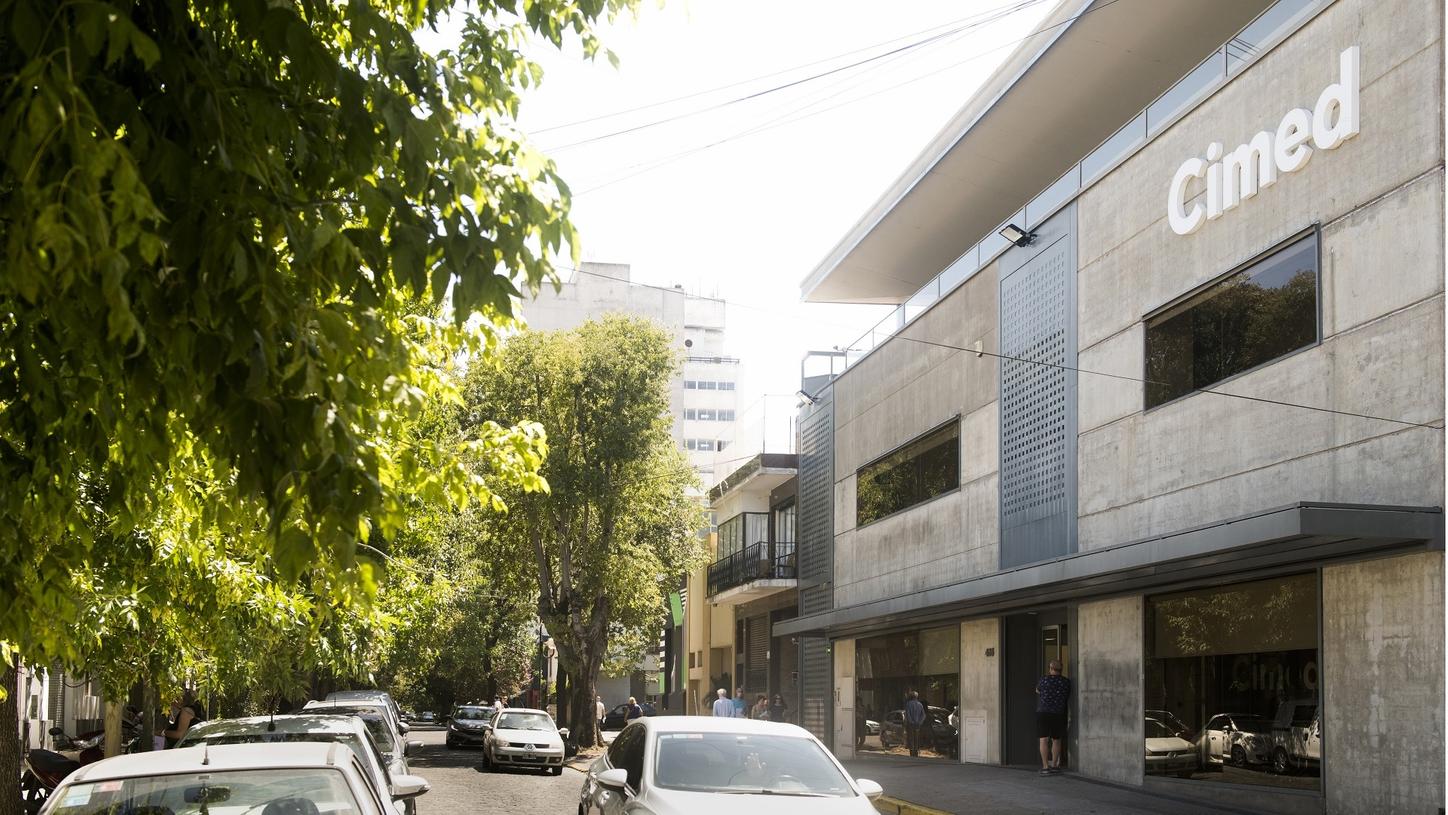
"Of course, it is a ‘Formula One’ machine, but what made the most difference for us when choosing the PET/CT was the level of technical support available through our relationship with Siemens Healthineers,” explains Mauro Piatti, MD, director of Cimed, a diagnostics clinic in La Plata. His team has been working with Biograph Horizon for the past six months.
Keeping pace with new challenges
Piatti’s father, Gustavo Poggio, MD, led the founding of Cimed in 1991, and the clinic now receives about 15,000 patients a month for medical imaging, from ultrasound and bone densitometry to PET/CT. “The referral is
spontaneous—we are not associated with any hospital or medical group. Instead, many doctors choose to refer the patient to us,” says Poggio.
According to Dos Anjos, one of the key ways to improve the patient experience in PET/CT is by reducing acquisition times. “The mean total scan time used to be about 20 minutes, and now we are scanning our patients in 15 minutes. It may seem like a small difference, but one needs to take into account the fact that most of them are oncologic patients who are suffering from pain,” he explains. “Five minutes is actually a lot of time for them.”
Another advantage of short acquisition times, according to Dos Anjos, is that there is less need to repeat scans due to inadvertent movements of tired patients—mainly in the region of the head—during longer sessions. He estimates that his team is now able to complete about three whole-body scans per hour. “The shorter acquisition times, lower doses, and fewer repetitions of the scans all add up to produce a great improvement to our patient workflow.”
Importance of speed and image quality
“Speed is very important,” agrees Poggio, “and so is imaging quality.” The ability to detect smaller and smaller lesions has also been a marked improvement, says Dos Anjos. “I was skeptical about this before working with Biograph Horizon. Now we are performing some PSMA PET scans in which we are able to detect 5-millimeter lymph nodes with very sharp contours.” The ability to identify such small lesions derives from Biograph Horizon’s small LSO crystals and its True time-of-flight technology, enabling high spatial resolution and better signal-to-noise ratio than prior to using the technology.
“We were looking for a PET/CT system with time-of-flight technology, and Biograph Horizon was the perfect choice for us in terms of cost-benefit ratio.”
Patients in La Plata are also being scanned in 15 minutes or even less time, says Juan Cuesta, MD, a radiologist and Cimed’s medical director. He explains he and his colleagues do their utmost to wed this technological efficiency with the culture of personalized care that Cimed has always emphasized.
“One thing that we try to do differently here is to have the doctors always stay close to the patients, from the time they arrive until the end of the scan, asking detailed questions about their condition and explaining the process,” says Cuesta. “The patient is not left alone at any time, being accompanied warmly, respectfully—and that makes a huge difference,” adds Poggio. This approach has also been helpful when the need to scan pediatric patients arises. He adds that in most cases, the team has managed to do this without the need to anesthetize those patients.
Reduced radiation treatments
In Brazil, Dos Anjos’ team at Dasa has seen a marked reduction in the dosage of radioisotopes, such as fludeoxyglucose F 18 (FDG), in their routine exams. “We usually had to inject a dose with about 1.5 millicurie per kilogram of FDG to achieve good imaging. Now, we are down to about 0.8 to 1.0 millicuries—an almost 50% reduction,” he continues “Some of the oncologic patients are scanned every two to three months, so we are talking about a significant exposure to radiation. It’s great if you can find ways to reduce it.”
“One thing that we try to do differently here is to have the doctors always stay close to the patients, from the time they arrive until the end of the scan, asking detailed questions about their condition and explaining the process.”
Although PET/CT scans in both centers are mainly focused on oncological treatments, Biograph Horizon has shown its usefulness for other specialties as well, such as neurology. “Five months ago, we started to perform amyloid PET scans that are now commercially available in Brazil, and it’s amazing,” says Dos Anjos. “In the older population, it’s not that specific to detect Alzheimer’s disease, since about 30% of elderly people have a positive amyloid PET scan without having Alzheimer’s. Now it’s starting to become very useful in detecting the early stages of the disease in patients who are younger.”
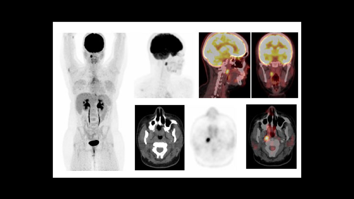
His Argentinian counterparts are doing similar work with Alzheimer’s and neurodegenerative diseases like Parkinson’s and epilepsy. According to Dos Anjos, there are also interesting possibilities to explore in cardiology. “The referral for endocarditis investigation is increasing, and it’s amazing how we can detect small defects in the heart.” The members of the La Plata team have also used their PET/CT capabilities to look for the causes of different medical conditions that have not been discovered by other methods.
Novel radioisotopes
Dos Anjos foresees exciting future research and clinical possibilities applying PET/CT, including the use of novel radioisotopes. “We are seeing a lot of clinical trials testing PSMA lutetium in association with other drugs in the earlier clinical stage of prostate cancer. PET images are being used more and more in these clinical trials, and I think they will be essential to patient selection and to treatment risk monitoring.”
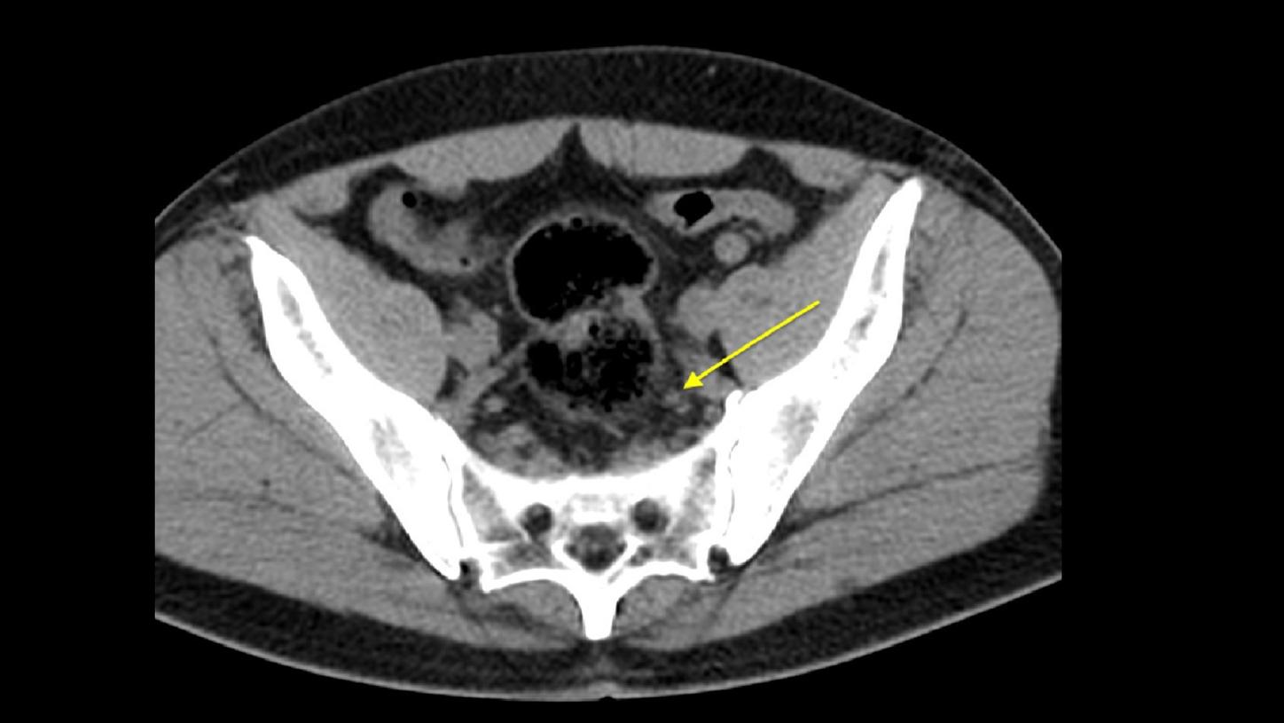
CT and PET/CT-fused axial images. A 59-year-old male patient with prostate adenocarcinoma underwent a PET/CT with 18F-PSMA-1007 for biochemical recurrence. PET/CT image showed a very small pelvic lymph node (5 mm) with high radiotracer uptake. Data courtesy of Dasa, São Paulo, Brazil.
Dos Anjos and his colleagues are also using Biograph Horizon to monitor treatments via radioembolization, using labeled microspheres. After the treatment, the patients receive a PET scan. “It’s a very nice way of making sure that the microspheres have been correctly delivered to tumors,” he explains. “Imaging is fundamental to monitor any treatment.”
About the Author
Reinaldo José Lopes is a science and health writer at Folha de S. Paulo, Brazil’s leading daily newspaper, and is the author of several books.
Fludeoxyglucose F 18
Brief summary and complete prescribing information for Fludeoxyglucose F 18 5-10mCi as an IV injection
Related Articles & More Information


