Syngo DynaCT
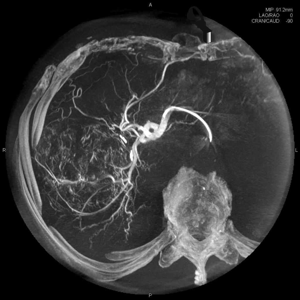


Syngo O DynaCT, lançado em 2004, é a solução de TC de feixe cônico da Siemens que permite aos usuários obter informações do tecido mole durante as intervenções. Ele ganhou reconhecimento como uma ferramenta altamente valiosa para vários procedimentos clínicos. Syngo O DynaCT melhora os resultados do paciente ao fornecer informações não aparentes no DSA e permite que os usuários ampliem o escopo do procedimento. Sendo percebido no passado como mais adequado para usuários experientes, o novo PURE ® plataforma agora torna mais fácil para todos os usuários obter resultados consistentes de alta qualidade.
- Suporte a resultados consistentes em TC de feixe cônico
- Melhore os resultados do paciente e evite complicações
- Amplie o escopo do procedimento do seu laboratório de angio
- Leia mais detalhes
Características e Benefícios
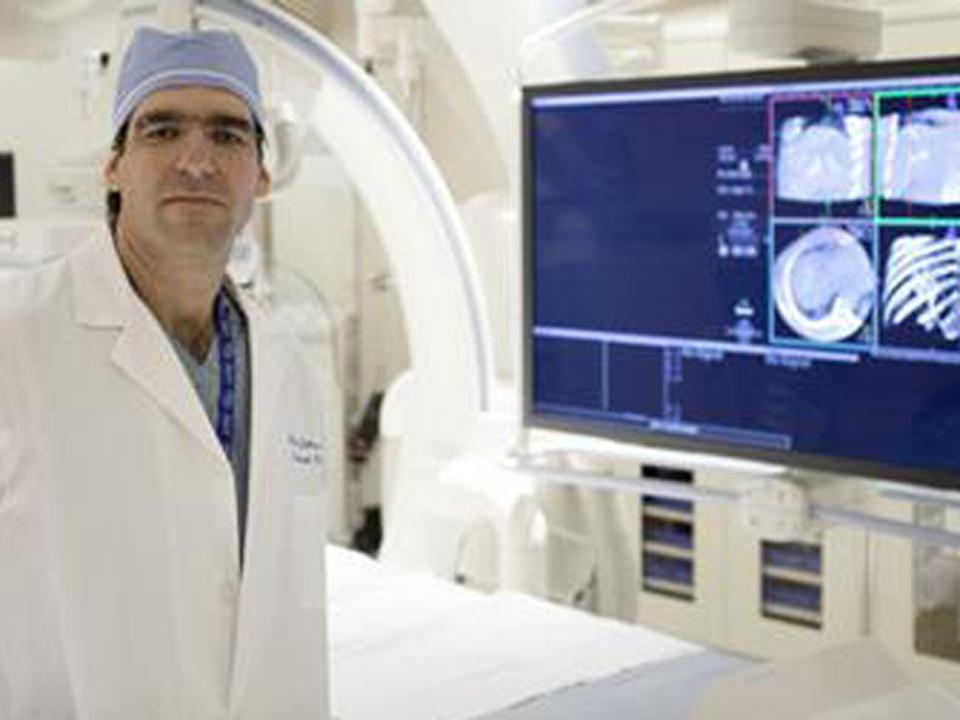
syngo DynaCT enables CT-like cross-sectional imaging by creating 3D soft tissue data sets. syngo DynaCT can be performed directly in the angio suite without loss of time and additional risk to the patient.
Ensure consistent results in cone-beam CT
In the past, different levels of expertise may sometimes have led to varying results when using cone-beam CT. In contrast, with the new 3D Wizard image-guided protocol selection, available on Artis with PURE®, syngo DynaCT is also accessible for occasional users. The 3D Wizard leads all users to consistent and high-quality results. It helps users select the correct protocol with a visual support of clinical images, offers all relevant information on delay, injection and patient positioning, and guides users through the acquisition, step by step. This supports standardized quality of care, regardless of who performs the procedure, from occasional to experienced users.
Improve patient outcome
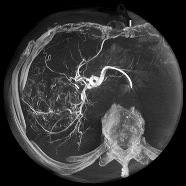
syngo DynaCT can provide information not apparent in DSA. This additional information may result in changes in diagnosis, treatment planning, or treatment delivery. It supports sounder decision-making during the intervention – for example when employed to exclude non-target embolization during liver tumor embolization or to confirm device position within intracranial vessels. Where in the past it would have been necessary to transfer the patient into the CT unit, syngo DynaCT can be performed instead, directly in the angio suite without loss of time and with no additional risk to the patient.

Broaden the procedure scope of your angio lab
Dedicated syngo DynaCT applications allow users to extend the clinical capabilities of their angio lab. syngo DynaCT Micro, for instance, enables users to visualize the smallest details, bringing additional procedures such as arthrography, intra-cranial stent placement follow-up examinations, or validation of cochlear implant placement to the angio suite. Other dedicated applications that further expand the confidence and performance of syngo DynaCT include syngo DynaCT SMART (Streak Metal Artifact Reduction Technique): This special technique, available for Artis with PURE®, substantially reduces metal artifacts, enabling sounder detection of bleeds even around small coil packages. syngo DynaCT 360 or syngo DynaCT Large Volume: Enable acquisition of images with an even larger volume, which can be very useful during liver embolization or needle procedures.

syngo DynaCT also enables users to take on recently developed, innovative procedures – thus extending their clinical capabilities even further. For instance, in new embolization procedures such as prostate artery embolization for treatment of benign prostatic hyperplasia, syngo DynaCT helps operators to clearly distinguish between target and non-target embolization sites. syngo DynaCT gives users the right tools to tackle exciting new prospects.
Uso Clínico
Siemens was first to introduce soft tissue imaging in the interventional suite as a breakthrough innovation for neuro applications. Continuous development in collaboration with institutions worldwide has made syngo DynaCT available for applications in the entire body. What started as a breakthrough is now a worldwide trend in interventional imaging. As of June 2015, hundreds of institutions around the world have installed syngo DynaCT in their angio suite.
"Our experience with syngo DynaCT is extremely positive. ... It has an incredibly high impact on our daily practice."1
Saruhan Cekirge, MD, Hacettepe University, Ankara, Turkey
"syngo DynaCT helped us to reduce the time for liver tumor treatment by 40%."1
Dr. Waggershauser, University of Munich, Germany
Expert statement
Professor Thomas J. Vogl, M.D.
Head of Diagnostic and Interventional Radiology University of Frankfurt - Germany
Case Studies

Minimally Invasive Spinal Osteosynthesis
An 88-year-old female patient suffered from lower back pain. She was operated on before due to a traumatic fracture of L5 which required cementoplasty.

Stent Placement across Cerebral Aneurysm
65-year-old male with hypertension and history of myocardial infarction and coronary stenting with incidental left internal carotid artery aneurysm.
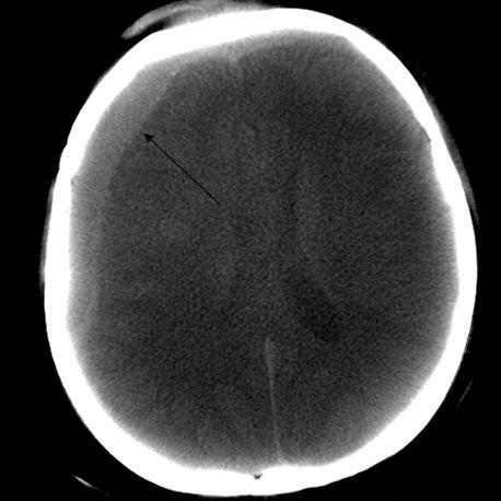
Cerebral trauma
Female patient with brain trauma for 3 days, mental imbalance and loss of consciousness.
Protocols and Sequences

syngo DynaCT in multiple phases for HCC
A 69-year-old male with HCC (hepatocellular carcinoma) and HCV (Hepatitis C virus)-positive hepatic cirrhosis.

Follow-up after stent-assisted aneurysm coiling
73-year-old female patient. Left middle cerebral artery (MCA) aneurysm that was treated with stent-assisted coil embolization in 2005. Because of her history of contrast allergies, we decided to complete a syngo DynaCT run with IV injection...

syngo DynaCT of the pulmonary arteries
Patient with history of recurrent pulmonary embolism and chronic thromboembolic pulmonary hypertension (CTEPH). Pulmonary angiogram and syngo DynaCT were acquired for diagnostic work-up.
Articles and Whitepapers
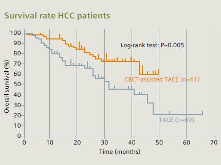
Use of Cone Beam CT (CBCT) in the angio suite improves safety and outcomes in interventional tumor treatment.
Dr. Michael J. Wallace on the Impact of Improved Image Quality of Angiography Equipment in Interventional Oncology.
Scientific Talks and Publications
General IR
- Transjugular Intrahepatic Portosystemic Shunt Creation in a Polycystic Liver Facilitated by Hybrid Cross-sectional/Angiographic Imaging (Daniel Y. Sze et al., JVIR 2006)
- Intra-operative DynaCT improves technical success of endovascular repair of abdominal aortic aneurysms (Lukla Biasi, MD et al., Journal of Vascular Surgery 2009)
- Comparison of C-arm Computed Tomography and Digital Subtraction Angiography in Patients with Chronic Thromboembolic Pulmonary Hypertension (Jan B. Hinrichs et al., CardioVascular and Interventional Radiology 2015)
- C-arm Cone-beam CT: General Principles and Technical Considerations for Use in Interventional Radiology (Robert C. Orth et al., Journal of Vascular and Interventional Radiology 2008)
Interventional Oncology
- Impact of 3D Rotational Angiography on Liver Embolization Procedures: Review of Technique and Applications (Pierleone Lucatelli et al., CardioVascular and Interventional Radiology 2015)
- Utility of C-arm CT in Patients with Hepatocellular Carcinoma undergoing Transhepatic Arterial Chemoembolization (Alessia Tognolini et al., Journal of Vascular and Interventional Radiology 2010)
Structural Heart Disease
- Using DynaCT for the assessment of iliofemoral arterial calibre, calcification and tortuosity index in patients selected for trans-catheter aortic valve replacement (James A. Crowhurst et al., The International Journal of Cardiovascular Imaging 2013)
- Impact of optimising fluoroscopic implant angles on paravalvular regurgitation in transcatheter aortic valve replacements – utility of three-dimensional rotational angiography (Karl K. Poon et al., Euro Intervention)
- Integration of MDCT and Fluoroscopy Using C-arm Computed Tomography to Guide Structural Cardiac Interventions in the Cardiac Catheterization Laboratory (Amar Krishnaswamy et al., Catheterization and Cardiovascular Interventions 2015)
- Effect of body mass index on the image quality of rotational angiography without rapid pacing for planning of transcatheter aortic valve implantation: a comparison with multislice computed tomography (Carl J. Schultz et al., European Heart Journal 2013)
- Transcatheter Aortic Valve Replacement using DynaCT (Koichi Maeda et al., Journal of Cardiac Surgery 2012)
- System to Guide Transcatheter Aortic Valve Implantations Based on Interventional C-Arm CT Imaging (Matthias John et al., Medical Image Computing and Computer-Assisted Intervention – MICCAI 2010)
- Multimodality Image Guidance With Dyna-CT for Transcatheter Treatment of Paravalvular Leak of a Stentless Valve (Brett Van Leer-Greenberg et al., Catheterization and Cardiovascular Interventions 2015)
Detalhes técnicos
How does syngo DynaCT work?
syngo DynaCT utilizes images acquired from a special rotational angiography run to reconstruct cross-sectional soft tissue information. Image acquisition is achieved in 5-20 seconds, depending on the protocol used. In that time the C-arm covers an angle of at least 200° and acquires the needed projection images for reconstruction. These images are automatically sent and reconstructed on a syngo X Workplace and available for assessment in the interventional suite.
What does “soft tissue information” mean?
Before the introduction of syngo DynaCT, 3D angiography enabled the visualization of high-contrast objects such as stents, clips, bones and contrast-filled vessels during interventional procedures. With syngo DynaCT, it is possible to achieve a higher level of tissue differentiation in the reconstructed images, enabling the visualization of objects with lower Hounsfield Units (HU) values.

With syngo DynaCT we can visualize objects of 5 HU and 10 mm in size or 10 HU and 5 mm in size*. This enables visualization of lower contrast objects such as larger hemorrhages and tumors.
* as measured using a 16-cm CATPHAN phantom
What is the FOV (field of view)?
syngo DynaCT datasets acquired with a ceiling, floor or biplane stands provide a volume of 18.5 cm height with a diameter of 24 cm.
The Artis zeego enables in addition larger volume acquisitions such as syngo DynaCT Large Volume and syngo DynaCT 360, which result in a volume of 18 cm height with a 47 cm diameter, or 24 cm height with a 35 cm diameter.

If desired, it is also possible to acquire a smaller volume using collimation* in order to reduce dose and increase image quality.
*collimation on the longitudinal access.
What is the spatial resolution ofsyngo DynaCT and what slice thickness can be reconstructed?
The slice thickness that can be reconstructed depends on the volume of interest and the voxel size. The standard slice thickness is about 0.5 mm and there are additional reconstruction options that allow creation of lower slice thickness. The spatial resolution achieved with syngo DynaCT is about 0.22 mm. In addition, with syngo DynaCT Micro, a special protocol is available that provides high resolution 3D imaging, boosting the level of detail by using each detector pixel. With syngo DynaCT Micro an even higher spatial resolution of about 0.14 mm can be achieved.
Does syngo DynaCT offer a dedicated body protocol?
Yes, syngo DynaCT offers a dedicated body protocol, about 400 projections are acquired in less than 6 seconds*. Due to its fast rotation this protocol reduces motion artefacts providing excellent image quality. High resolution results (512 matrix) are available at tableside in less than 15 seconds*. In addition there is a dedicated syngo DynaCT protocol with reduced dose and shorter acquisition time for dose-sensitive applications.
*Mentioned values are for Artis with PURE and may change depending on system hardware and specific protocol used.
Does syngo DynaCT offer a dedicated neuro protocol?
Yes, there are several syngo DynaCT protocols that can be useful for interventional neuroradiology procedures.
The standard dedicated neuro protocol acquires about 500 projections in 20 seconds. High resolution results (512 matrix) are available at tableside in about 30 seconds*.
There are also protocols for reconstructing information of subtracted scans enabling the easy visualization of vessels in relation to devices such as stents or coil packages and a special protocol for stent follow-up using an intravenous injection.
In addition, with syngo DynaCT SMART (Streak Metal Artifact Reduction Technique) a special reconstruction is included for reducing metal artifacts in the resulting images, which increases the diagnostic confidence and the chance to visualize complications such as bleeds close to metallic objects.
* Mentioned values are for Artis with PURE and may change depending on system hardware and specific protocol used.
General Requirements
System
- ARTIS pheno
- Artis Q
- Artis Q.zen
- Artis zeego
- Artis zee
- Artis zeego
Minimum Software Version
syngo X Local de Trabalho: VB21
Other
Por favor, observe: pré-requisitos técnicos adicionais podem ser aplicados. Ao receber sua solicitação, seu representante local da Siemens Healthineers esclarecerá se o seu sistema atende aos requisitos.