In the highly competitive healthcare environment, differentiation is key to maintaining competitiveness. With the Biograph mMR PET-MR system you can set the pace in diagnostic imaging, by combining state-of-the-art 3T MRI with proven molecular imaging, fully integrated in one system. With syngo MR E11, we take hybrid imaging to the new level of Synergistic PET-MR to significantly improve diagnostic accuracy, capabilities as well as the standardization of quality of care – for both research and clinical use.

Biograph mMRFrom simultaneous to synergistic PET-MR
Features & Benefits
Biograph mMR has revealed additional findings and led to a change in therapy in 18%1 of oncologic staging and follow-up patients. With syngo MR E11, we enhance an already leading Synergistic PET-MR. Now, you can improve diagnostic accuracy and standardize quality.

Benefit from motion-free PET images with MR-based motion compensation beyond gating
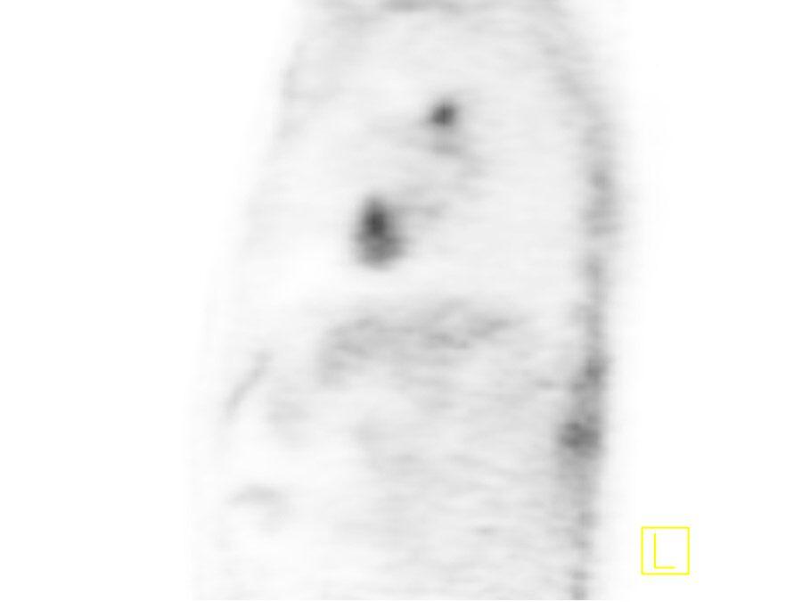
Cancer rates are increasing globally. Over 14 million adults were diagnosed with cancer in 2012.2 At the same time treatment cost can become excessive.3 Biograph mMR is the key to more accurate diagnostic accuracy - for example with the Siemens' unique BodyCOMPASS. This technology enables motion-free PET images with MR-based motion compensation beyond gating, also in challenging body regions like the abdomen.
MR-based motion compensation with BodyCOMPASS
 Static image
 Motion corrected image
NYU, Langone Medical Center,
New York, USA
MR-based motion compensation delivers sharp delineation of lung lesion, with accurate SUV uptake.

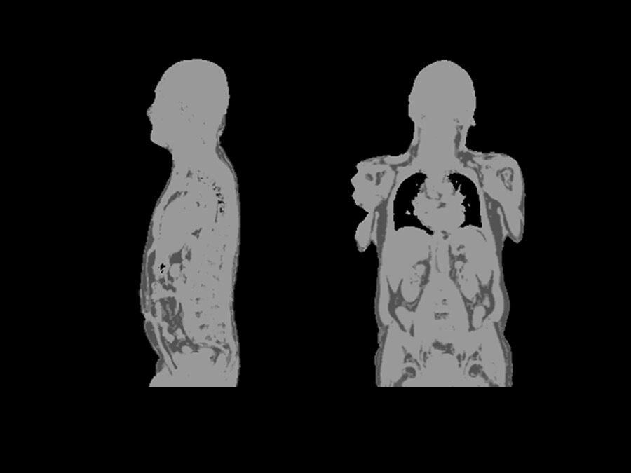
Advance PET attenuation correction with whole-body 5-compartment model including bones and HUGE
Metabolic information from PET adds important data to the morphological and anatomical information from the MR. So far only air, fat, water, and tissue were included. Now, by including all major bones into the patient attenuation map, the evaluation of lesions in and close to bones is improved. Improve diagnostic capabilities with the unique whole-body 5-compartment model to reach the new level of Synergistic PET-MR.
Discover the unique whole-body 5-compartment attenuation correction model including bones


University Hospital Erlangen,
Erlangen Germany
Now, with unique 5-compartment attenuation correction model including all major bones you are able to better delineate lesions and improve diagnostics capabilities.
Improve diagnostic capabilities with these solutions:
- Overcome truncations in the arms by extending the FoV of MR to the edges of the bore with HUGE
- Achieve independence from tracer by utilizing fully MR-based attenuation correction

Deliver exceptional quality and speed in PET-MR with the latest MR innovations
Diverse requests from referrers and varying compliance of patients such as children4 or very ill patients places further challenges on an already competitive healthcare environment. With the latest software platform syngo MR E11, Biograph mMR helps answer these demands and provides a consistent and standardized quality of care.
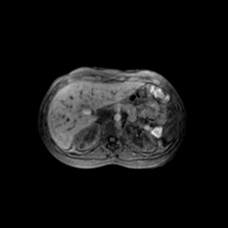
FREEZEit - embrace motion in liver imaging
 conventional
 StarVIBE
University Hospital Grosshadern,
Munich, Germany
Standardize quality of care with syngo MR E11:
Clinical Use


University Hospital Grosshadern, Munich, Germany

St. Bartholomew`s Hospital, London, United Kingdom


University Hospital Grosshadern, Munich, Germany

St. Bartholomew`s Hospital, London, United Kingdom


Technical Details
We've designed the first PET detectors that allow the integration of whole-body MR and PET - without compromising the performance of either modality.
Magnet System |
|
Field strength | 3 Tesla |
Bore size | 60 cm / 1.97 ft |
Magnet length (cover to cover) | 199 cm / 6.53 ft |
Helium consumption | Zero Helium boil-off technology |
Shimming | Passive and active; Automatic, patient specific shim; 3 linear and 5 non-linear (2nd order) |
|
|
Gradient strength | MQ Gradients (45 mT/m @200 T/m/s) |
|
|
RF technology |
|
Maximum number of channels5 | 102 |
Number of independent receiver channels that can be used simultaneously in one single scan and in one single FoV, each generating an independent partial image | 18, 32 |
|
|
Siting and Installation |
|
System length incl. patient table | 335 cm / 10.99 ft |
System weight (in operation) | 9 tons / 19,842.60 lb |
Power requirement | 110 kVA connection |
Minimum room size6 | 33 m2 / 355 sq ft |
|
|
PET Detectors |
|
Crystal material | LSO |
Crystal element dimension | 4 mm x 4 mm x 20 mm |
|
|
Transverse spatial resolution (NEMA 2012) |
|
FWHM @ 1 cm | 4.6 mm |
Sensitivity | 14.1 cps/kBq |
Peak NEC rate (at ≤26 kBq/cc) | 180 kcps |
Axial FoV for PET | 258 mm |
Initial Experience in 134 Patients—A Hypothesis-generating Exploratory Study; Radiology, 2013.


