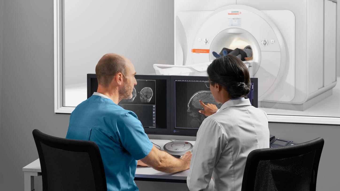
Explore new territories in MRI by enabling powerful 7T research and enhancing clinical care and better patient outcomes. MAGNETOM Terra is the first 7T scanner released for clinical use in Europe and the US.
The unique Dual Mode lets you switch between clinical and research operations, with separate databases to distinguish between clinical and research scans.1
Our advanced ultra-high-field (UHF) technology will keep you at the cutting edge of MRI, to attract the brightest minds to your facility, sharpen your competitive edge and strengthen your reputation.

MAGNETOM TerraTranslate 7T research power into clinical care
Features & Benefits
The power to reach further and enter new territories is essential for those who shape the future of healthcare. Whether for basic, clinical or neuroscience research endeavors, MAGNETOM Terra lets you go deeper than ever before. All in one, the power of 7T MRI helps you deliver previously unseen insights that could improve patient outcomes and further enhance your reputation.

Double SNR for more precision2 – With clinical applications in Dual Mode
When it comes to improving patient outcomes by answering the most challenging clinical questions, enabling deeper insights can make a difference. With double the SNR of 3T, imaging at 7T offers potential higher resolution in both anatomical and functional imaging.

Help improve patient outcomes:
- First 7T scanner released for clinical use
- 0.2 mm in-plane resolution to visualize previously unseen structures
- 0.14 cm³ voxel sizes for innovative metabolic brain mapping 3
- Submillimeter BOLD fMRI precision to visualize sub-cortical activations4

Unlock research beyond clinical limits – With up to 16-channel parallel transmit1
When it comes to research, the freedom to explore new territories is imperative in order to gain a competitive edge. MAGNETOM Terra unlocks a whole new world of potential for basic, clinical and neuroscience research helping you translate your outcomes into better clinical care. The parallel transmit technology of MAGNETOM Terra with up to 16-channel pTX gives you all the freedom you need for the imaging of challenging body regions.

Unlock powerful research capabilities:
- Visualize sub-cortical activations with BOLD fMRI4
- More power for diffusion MRI with 80/200 gradients
- Higher acceleration factors with 64 receive channels5

Physiology is at your fingertips
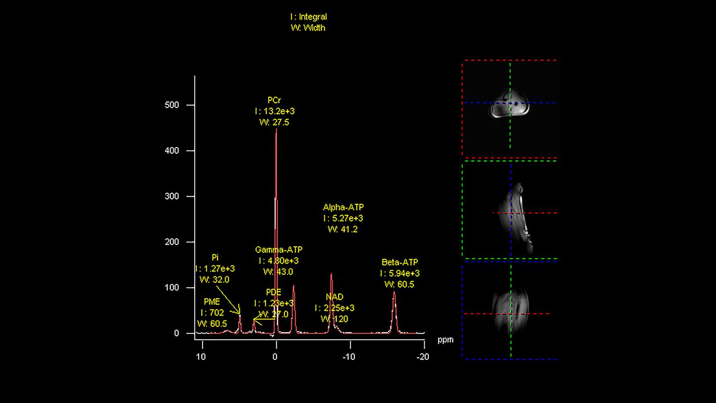
MAGNETOM Terra is the first 7T MRI scanner to unleash the full potential of the increased MR signal with multinuclear imaging and spectroscopy in clinical settings. The multinuclear option allows the use of two dedicated coils in clinical mode – a 23Na head coil and a 31P loop coil, to explore metabolic insights. In research mode, MAGNETOM Terra enables the use of up to 10 nuclei.6

Explore physiology with multinuclear MR:
- Sodium MRI and phosphorus MRS in clinical mode
- Dedicated coils for multinuclear applications
- Support of up to 10 nuclei in research mode1

Change the game in UHF business – With Siemens’ 50% lighter 7T magnet7
MAGNETOM Terra provides leading-edge capabilities to increase your competitiveness, while making a tangible difference to clinical care, research – and your business. The new Siemens 50% lighter 7T magnet7 is designed to enable easier integration into clinical environments and lower operating costs.

The right business impact:
- Easy integration in clinical environments, faster installation
- Reduced operating costs thanks to Zero Helium boil-off
- All of MAGNETOM Terra's key components are delivered from a single reliable partner you can trust

Join the largest research community – With over 75% of all UHF users
Your reputation plays a pivotal role in your institution’s success. MAGNETOM Terra helps you attract the brightest minds to your facility, to consequently publish first and put your organization firmly on the map. With over 75% of all UHF scanners deployed worldwide, Siemens fosters access to an exclusive network of expertise and broad scope for collaboration and exchange.

Increase your reputation:
- Over 75% of 7T MRI scanners worldwide are from Siemens
- In 2018, more than 73% of UHF ISMRM abstracts were based on data on Siemens Healthineers scanner.8
Clinical Use
In order to set new trends, it is crucial to overcome technical and field strength related limitations in MRI. MAGNETOM Terra has the power to let you delve deeper than ever before, making your research stand out from the rest with the potential to improve patient outcomes.
Watch Professor Siegfried Trattnig, MD, MedUni of Vienna, explain what makes imaging at 7T an important step for future clinical routine applications9:
Chemical Exchange Saturation Transfer (CEST)
Similar sodium MR images, CEST imaging provides also glycosaminoglycans information, but with higher image resolution. First studies show potential for detecting early stages of osteoarthritis.
Ultra-high resolution imaging of the hippocampus
In vivo visualization of the hippocampal sub-structures is now possible with 7T MRI. 0.2 mm in-plane resolution offers potential for clinical evaluation in patients suffering from temporal lobe epilepsy and Alzheimer’s disease.
Presurgical fMRI in oncology
Ultra-high resolution fMRI in the brain sensor motor area offers potential for more accuracy in the presurigcal evaluation of tumor patients.
Ultra-high resolution venography
Susceptibility Weighted Imaging (SWI) in oncology can help differentiate between high and low grade benign and malignant tumors in the brain by depicting previously unseen information.
Phase contrast in Multiple Sclerosis
Standard FLAIR together with SWI at 7T may help in better understanding Multiple Sclerosis. Detecting iron accumulation and the vein density in plaques can be possible thanks to ultra-high resolution at 7T.
Metabolic brain mapping
Proton MR spectroscopy at 7T can be used to provide not only metabolic information, but also anatomical information. Ultra-high resolution spectroscopy has potential for clinical applications such as in tumors, epilepsy, Multiple Sclerosis and neurodegenerative diseases.
MSK morphology imaging
High resolution imaging offers potential for clinical applications in orthopedics where the details make a difference. Knee, ankle, hand, and foot imaging of joints and cartilage help improve diagnostics, for example, in osteoporosis.
High-resolution cartilage imaging
High-resolution T2 mapping and non-contrast sodium imaging for the quantification of glycosaminoglycans offer potential for the assessment of biochemical and biomechanical information of transplant cartilage.
Chemical Exchange Saturation Transfer (CEST)
Similar sodium MR images, CEST imaging provides also glycosaminoglycans information, but with higher image resolution. First studies show potential for detecting early stages of osteoarthritis.

FID nonselective
TR = 400 ms, TE = 0.23 ms
256 averages
TA = 1:42 min
4aaaa0244 FAU, Erlangen, Germany

SWI MIP
0.2 × 0.2 × 1.0 mm3
TA = 10:57 min
4aaaa0055 FAU, Erlangen, Germany

TOF
4 mm isotropic
TA = 9:32 min
4aaaa0116 FAU, Erlangen, Germany

T2 TSE
0.4 × 0.4 × 3.0 mm3
TA = 5:19 min
4aaaa0058 FAU, Erlangen, Germany

MP2RAGE
0.7 mm isotropic
TA = 13:40 min
4aaaa0136 FAU, Erlangen, Germany

23Na UTE
3 mm isotropic
TA = 4:08 min
Fused with TSE
4aaaa0243 FAU, Erlangen, Germany

PD TSE fs
0.4 × 0.4 × 2.5 mm3
TA = 3:00 min
4aaaa0066 FAU, Erlangen, Germany

3D DESS we
0.5 mm
TA = 3:41 min
4aaaa0250 FAU, Erlangen, Germany

FID nonselective
TR = 400 ms, TE = 0.23 ms
256 averages
TA = 1:42 min
4aaaa0244 FAU, Erlangen, Germany

SWI MIP
0.2 × 0.2 × 1.0 mm3
TA = 10:57 min
4aaaa0055 FAU, Erlangen, Germany

TOF
4 mm isotropic
TA = 9:32 min
4aaaa0116 FAU, Erlangen, Germany

T2 TSE
0.4 × 0.4 × 3.0 mm3
TA = 5:19 min
4aaaa0058 FAU, Erlangen, Germany

MP2RAGE
0.7 mm isotropic
TA = 13:40 min
4aaaa0136 FAU, Erlangen, Germany

23Na UTE
3 mm isotropic
TA = 4:08 min
Fused with TSE
4aaaa0243 FAU, Erlangen, Germany

PD TSE fs
0.4 × 0.4 × 2.5 mm3
TA = 3:00 min
4aaaa0066 FAU, Erlangen, Germany

3D DESS we
0.5 mm
TA = 3:41 min
4aaaa0250 FAU, Erlangen, Germany

FID nonselective
TR = 400 ms, TE = 0.23 ms
256 averages
TA = 1:42 min
4aaaa0244 FAU, Erlangen, Germany


Technical Details
MAGNETOM Terra comes equipped with the latest ultra-high-field technological features, aiding you in obtaining excellent image quality to support your 7T MRI clinical and research endeavors.
Magnet System |
|
Field strength | 7 Tesla |
Bore size | 60 cm |
Magnet length | 270 cm |
Helium consumption | Zero Helium boil-off technology |
Shimming | Passive and active |
|
|
Gradient strength | XR Gradients (80 mT/m @ 200 T/m/s) |
|
|
RF technology |
|
Maximum number of channels10 | 32, 64 |
Number of independent receiver channels that can be used simultaneously in one single scan and in one single FoV, each generating an independent partial image | 32, 64 |
|
|
Siting and Installation |
|
System length | 297 cm |
System weight (in operation) | < 25 tons |
Minimum room size11 | 65 m2 (w/o pTX and 3rd order shim option) |


