Siemens Healthineers is working to bring the benefits of CMR to an even broader range of patients with free breathing exams, highly automated CMR exams, MR Angiography and our dedicated cardiovascular MRI scanner. Take a look at the clinical gallery to find out what's possible with our applications.
- Home
- Medical Imaging
- Magnetic Resonance Imaging
- Clinical Fields
- Cardiovascular MRI

Cardiovascular MRITransform your CMR practice with cutting-edge applications
Highlights
3D WholeHeart Pro1
The 3D WholeHeart Pro addresses complex cardiovascular MR imaging needs, offering high isotropic resolution and 3D tissue characterization essential for evaluating small lesions and extensive tissue damage.
It incorporates advanced automation algorithms for virtually artifact-free images and accurate coronary artery visualization in free-breathing for patients who struggle with breath-holding. 3D WholeHeart Pro integrates AI-based automation tools like AutoPositioning and AutoRestingPhase, and AutoTI streamlining the imaging process and ensuring high diagnostic quality.
AutoMate Cardiac1
AutoMate Cardiac enhances cardiovascular MR imaging by using AI-based, automated workflows to set up scan parameters, ensuring robust image quality regardless of operator expertise.
AutoMate Cardiac standardizes the imaging process, reduces complexity, and minimizes errors and redo scans. With features like AutoPositioning, AutoRestingPhase detection, and AutoTI proposal. AutoMate Cardiac optimizes CMR exams making them more efficient and reproducible.
BioMatrix Beat Sensor for CMR exams without ECG2
BioMatrix Beat Sensor enables users to perform CMR exams without ECG. This break-through technology uses the heart's own pulsile motion to trigger the exam. The result is faster CMR exams and improved patient experience.
Ever wonder how break-through technology is developed?
Cardiovascular applications
Cardiovascular magnetic resonance (CMR) imaging is the gold standard for evaluating myocardial function, volumes, and scar. CMR is unique in its ability to provide tissue characterization for differential diagnosis of myocardial disease. Recent guidelines also include indications for CMR in congenital heart disease, heart failure, and coronary artery disease (CAD).3
Free breathing exams
3D WholeHeart Pro1
The 3D WholeHeart Pro addresses complex cardiovascular MR imaging needs, offering high isotropic resolution and 3D tissue characterization essential for evaluating small lesions and extensive tissue damage.
It incorporates advanced automation algorithms for virtually artifact-free images and accurate coronary artery visualization in free-breathing for patients who struggle with breath-holding. 3D WholeHeart Pro integrates AI-based automation tools like AutoPositioning and AutoRestingPhase, and AutoTI streamlining the imaging process and ensuring high diagnostic quality.

Compressed Sensing Cardiac Cine
Compressed Sensing enables diagnostic imaging quality, even for patients with arrhythmias and those unable to hold their breath.
It supports segmented Cine imaging, offering high spatial and temporal resolution in brief breath-holds. Additionally, the Compressed Sensing real-time technique ensures high-quality imaging during free-breathing. These advancements significantly accelerate cardiac scans, boost efficiency, and broaden the range of patients eligible for Cardiac MRI.

PSIR HeartFreeze4
HeartFreeze, with motion compensation algorithms, enables high-resolution Late Gadolinium Enhancement (LGE) imaging in free-breathing.
This technology extends the benefits of detailed viability assessments to patients with arrhythmias or those unable to hold their breath.
Highly efficient, automated CMR exams
AutoMate Cardiac1
AutoMate Cardiac enhances cardiovascular MR imaging by using AI-based automated workflows to set up scan parameters, ensuring robust image quality regardless of operator expertise.
With features like planned AutoPositioning, AutoRestingPhase detection, and AutoTI proposal, AutoMate Cardiac standardizes the imaging process, reduces complexity, and minimizes errors and redo scans.
myExam Cardiac Assist
myExam Cardiac Assist is a unique offering from Siemens Healthineers which enables comprehensive Cardiac MR examinations in under 30 minutes.
This highly automated tool leverages AI-powered algorithms like AutoAlign to facilitate automatic planning of all cardiac views, ensuring standardized and reproducible cardiac scans. With its step-by-step guidance, myExam Cardiac Assist simplifies the complexities of cardiac exams. It can be used alone or in combination with AutoMate Cardiac1.

BioMatrix Beat Sensor2
BioMatrix Beat Sensor enables users to perform CMR exams without ECG. This break-through technology uses the heart's own pulsile motion to trigger the exam. The result is faster CMR exams and improved patient experience.
Dedicated CMR appliactions

High Bandwidth Inversion Recovery
High Bandwidth Inversion Recovery (HBIR) significantly improves the quality of CMR imaging in patients prone to susceptivity artefacts. This advanced sequence employs wideband techniques to minimize distortions, which enhances tissue characterization for optimal imaging of myocardial injuries.
HBIR is particularly valuable as it extends high-quality diagnostic capabilities to patients prone to metal artefacts and thereby expands the potential for accurate disease management and diagnosis.

MyoMaps
MyoMaps facilitates pixel-based quantification of myocardial tissue, aiding in the differential diagnosis of myocardial diseases. When used in conjunction with HeartFreeze, our motion correction algorithm, MyoMaps enables the inline generation of T1, T2, and T2* colored parametric maps.

4D Flow
Flow quantification provides crucial insights into cardiac hemodynamics and function. ECG-triggered 2D phase contrast imaging enables non-invasive, quantitative assessment of blood flow. With retrospective reconstruction, full heart cycle coverage is achieved, allowing comprehensive analysis.
Our enhanced 4D Flow acquisition offers complete volumetric coverage, significantly improving the orientation and visualization of blood flow and velocity throughout the heart and thorax. This technical advancement facilitates the detailed analysis of key hemodynamic parameters over the entire cardiac cycle.
MR Angiography
myExam Angio Assist
myExam Angio Assist facilitates the acquisition of optimally timed contrast images through interactive bolus timing.
This technology ensures consistent timing of the contrast agent for each angiography examination. It enables reliable imaging results, providing precise information to referring physicians and ensuring consistent, optimized kinetic timing for reading and reporting MR angiography cases.

QISS
QISS technology provides an accurate non-contrast alternative for visualizing arterial disease, especially in patients where contrast agents are contraindicated or those with renal dysfunction.
QISS MRA is an easy-to-use approach for imaging peripheral arteries and contributes to patient safety by reducing the need for contrast agents and the associated risks.
Our dedicated MRI Scanner

MAGNETOM Sola Cardiovascular Edition
Outcome relevant decisions – redefining patient pathways
MAGNETOM Sola Cardiovascular Edition automatically adjusts to patient biovariability to overcome unwarranted variations in cardiac MRI examinations:
- Free breathing exams with Compressed Sensing Cardiac Cine
- Tissue characterization with MyoMaps and HeartFreeze
- CMR for challenging patients with High Bandwidth Inversion Recovery
- Highly efficient CMR exams with BioMatrix Beat Sensor and myExam Cardiac Assist
Clinical gallery

Image courtesy Northwestern University Feinberg School of Medicine, Chicago, USA.
1 Hodnett-AJR-197-1466; 2 Knobloch-ClinRadiol-69-4854
QISS
QISS for non-CE peripheral MRAs in patients with renal insufficiencies. Accuracy comparable to contrast enhanced MRA.1,2
Courtesy of Evangelia Nyktari, M.D, Onassis Cardiac Surgery Center, Athens, Greece. Study ID: 0aaaa0022
3D WholeHeart Pro1
Quality thoracic imaging despite presence of devices
Image acquired with MAGNETOM Sola (1.5T)

Courtesy of Evangelia Nyktari, M.D.; Onassis Cardiac Surgery Center, Athens, Greece. Study-ID: 0aaaa0030
3D WholeHeart Pro1
LGE tissue characterization for Electrophysiology planning. Pre-procedural images loaded into electroanatomic mapping system.
Image acquired with MAGNETOM Sola (1.5T)

Images courtesy of Reza Hajhosseiny, Kings College London, UK. Study-ID: 0aaaa0023
3D WholeHeart Pro1
3D WholeHeart Pro for coronary artery visualization. Non-contrast CMRA compared to contrast-enhanced CCTA.
Image aquired with MAGNETOM Aera (1.5T)
Case courtesy: J. Dehem, Jan Yperman Ziekenhuis, Ypers, Belgium. Study-ID: 0aaaa04
Compressed Sensing & PSIR HeartFreeze4
Compressed Sensing Cardiac Cine and PSIR HeartFreeze4 for functional and LGE imaging in free breathing.
Image aquired with MAGNETOM Sola (1.5T)

Courtesy: St. Georg Hospital, Leipzig, Germany. Study-ID: 1aaaa2598
PSIR HeartFreeze4
PSIR HeartFreeze for LGE scar assessment in free-breathing
Image aquired with MAGNETOM Altea (1.5T)

Cases courtesy of Ch. Tillmanns, Diagnostikum, Berlin, Germany. Study-ID: 0aaaa26
MyoMaps
MyoMaps tissue characterization for the differential diagnosis of cardiomyopathies.
Image aquired with MAGNETOM Skyra (3T)

Courtesy of Dr. J. Dehem, Jan Yperman Hospital, Ieper, Belgium. Study-ID: 1aaaa4056
MyoMaps
MyoMaps provides pixel-based tissue quantification, in this case suggesting hypertrophic cardiomyopathy.
Image aquired with MAGNETOM Sola (1.5T)

Case courtesy of H. Vágó, Semmelweis Uni., Budapest, Hungary. Study-ID: 0aaaa0012
High Bandwidth Inversion Recovery
High Bandwidth Inversion Recovery for patients prone to susceptibility artefacts.
Image aquired with MAGNETOM Aera (1.5T)
Siemens Healthineers. Study-ID: 2aaaa1877
BioMatrix Beat Sensor2
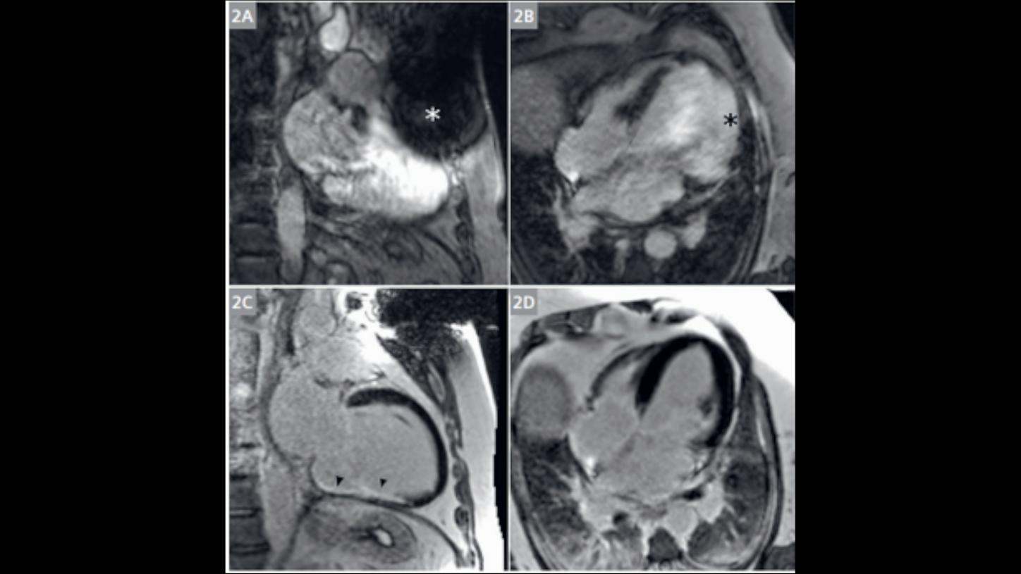
Comparison: Cardiac triggering with ECG versus cardiac triggering using Beat Sensor.
Image aquired with MAGNETOM Sola (1.5T)
Courtesy Dr. Johan Dehem, Jan Yperman Hospital, Ieper, Belgium. Study-ID: 1aaaa4048
BioMatrix Beat Sensor2
Cardiac Triggering without ECG.
Image aquired with MAGNETOM Sola (1.5T)
Study ID: 2aaaa1808
4D Flow imaging
4D Flow CMR imaging to quantify and visualize hemodynamics and morphology disorders of the heart.
Case courtesy: J. Dehem, Jan Yperman Ziekenhuis, Ypers, Belgium. Study-ID: 0aaaa0005
MR Angiography
Contrast enhanced axial T1FS blade imaging in carotid artery disease.
Image aquired with MAGNETOM Sola (1.5T)

Image courtesy Northwestern University Feinberg School of Medicine, Chicago, USA.
1 Hodnett-AJR-197-1466; 2 Knobloch-ClinRadiol-69-4854
QISS
QISS for non-CE peripheral MRAs in patients with renal insufficiencies. Accuracy comparable to contrast enhanced MRA.1,2
Courtesy of Evangelia Nyktari, M.D, Onassis Cardiac Surgery Center, Athens, Greece. Study ID: 0aaaa0022
3D WholeHeart Pro1
Quality thoracic imaging despite presence of devices
Image acquired with MAGNETOM Sola (1.5T)

Courtesy of Evangelia Nyktari, M.D.; Onassis Cardiac Surgery Center, Athens, Greece. Study-ID: 0aaaa0030
3D WholeHeart Pro1
LGE tissue characterization for Electrophysiology planning. Pre-procedural images loaded into electroanatomic mapping system.
Image acquired with MAGNETOM Sola (1.5T)

Images courtesy of Reza Hajhosseiny, Kings College London, UK. Study-ID: 0aaaa0023
3D WholeHeart Pro1
3D WholeHeart Pro for coronary artery visualization. Non-contrast CMRA compared to contrast-enhanced CCTA.
Image aquired with MAGNETOM Aera (1.5T)
Case courtesy: J. Dehem, Jan Yperman Ziekenhuis, Ypers, Belgium. Study-ID: 0aaaa04
Compressed Sensing & PSIR HeartFreeze4
Compressed Sensing Cardiac Cine and PSIR HeartFreeze4 for functional and LGE imaging in free breathing.
Image aquired with MAGNETOM Sola (1.5T)

Courtesy: St. Georg Hospital, Leipzig, Germany. Study-ID: 1aaaa2598
PSIR HeartFreeze4
PSIR HeartFreeze for LGE scar assessment in free-breathing
Image aquired with MAGNETOM Altea (1.5T)

Cases courtesy of Ch. Tillmanns, Diagnostikum, Berlin, Germany. Study-ID: 0aaaa26
MyoMaps
MyoMaps tissue characterization for the differential diagnosis of cardiomyopathies.
Image aquired with MAGNETOM Skyra (3T)

Courtesy of Dr. J. Dehem, Jan Yperman Hospital, Ieper, Belgium. Study-ID: 1aaaa4056
MyoMaps
MyoMaps provides pixel-based tissue quantification, in this case suggesting hypertrophic cardiomyopathy.
Image aquired with MAGNETOM Sola (1.5T)

Case courtesy of H. Vágó, Semmelweis Uni., Budapest, Hungary. Study-ID: 0aaaa0012
High Bandwidth Inversion Recovery
High Bandwidth Inversion Recovery for patients prone to susceptibility artefacts.
Image aquired with MAGNETOM Aera (1.5T)
Siemens Healthineers. Study-ID: 2aaaa1877
BioMatrix Beat Sensor2
Comparison: Cardiac triggering with ECG versus cardiac triggering using Beat Sensor.
Image aquired with MAGNETOM Sola (1.5T)
Courtesy Dr. Johan Dehem, Jan Yperman Hospital, Ieper, Belgium. Study-ID: 1aaaa4048
BioMatrix Beat Sensor2
Cardiac Triggering without ECG.
Image aquired with MAGNETOM Sola (1.5T)
Study ID: 2aaaa1808
4D Flow imaging
4D Flow CMR imaging to quantify and visualize hemodynamics and morphology disorders of the heart.
Case courtesy: J. Dehem, Jan Yperman Ziekenhuis, Ypers, Belgium. Study-ID: 0aaaa0005
MR Angiography
Contrast enhanced axial T1FS blade imaging in carotid artery disease.
Image aquired with MAGNETOM Sola (1.5T)

Image courtesy Northwestern University Feinberg School of Medicine, Chicago, USA.
1 Hodnett-AJR-197-1466; 2 Knobloch-ClinRadiol-69-4854
QISS
QISS for non-CE peripheral MRAs in patients with renal insufficiencies. Accuracy comparable to contrast enhanced MRA.1,2


Not for sale in the U.S.
