Reduce door-to-needle times in stroke
Computed Tomography can enhance clinical capabilities by providing better diagnostic confidence and improving process efficiency by saving working steps and making entire patient pathways even faster. Since stroke is the second leading cause of death worldwide, we support our customers in getting further with CT Neuro imaging.

NeurologyDriving progress to reduce door-to-needle time in stroke.
Features & Benefits
Reduce door-to-needle times
CT helps you quickly answer the most crucial diagnostic questions in stroke – so you can concentrate on optimal treatment and therapy decisions.
The three most crucial diagnostic questions in stroke
- Is the stroke caused by bleeding?
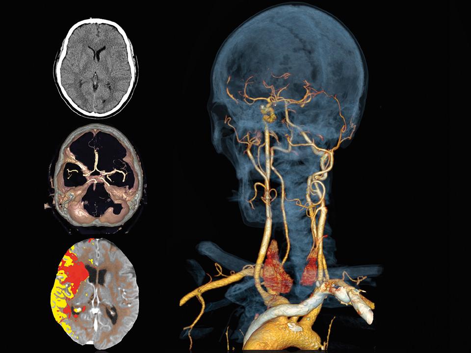
Use a non-contrast CT scan to detect if the stroke is caused by hemorrhage or ischemia, so you can determine the potential benefit or harm of thrombolytic drugs. Subtle nuances indicative of early signs of ischemia are visualized with Neuro BestContrast, which enhances the contrast-to-noise ratio by up to 40 percent.
- What is the size and location of the clot?
When planning interventional clot retrieval, the size of the clot may be overestimated on axial CTA source images. syngo.CT Dynamic Angio can help you better characterize the occlusion length and collateral status. Videos showing the flow of contrast from the arteries to the veins enable a dynamic evaluation so you can see antegrade and delayed collateral blood flow.
- How big is the infarct?
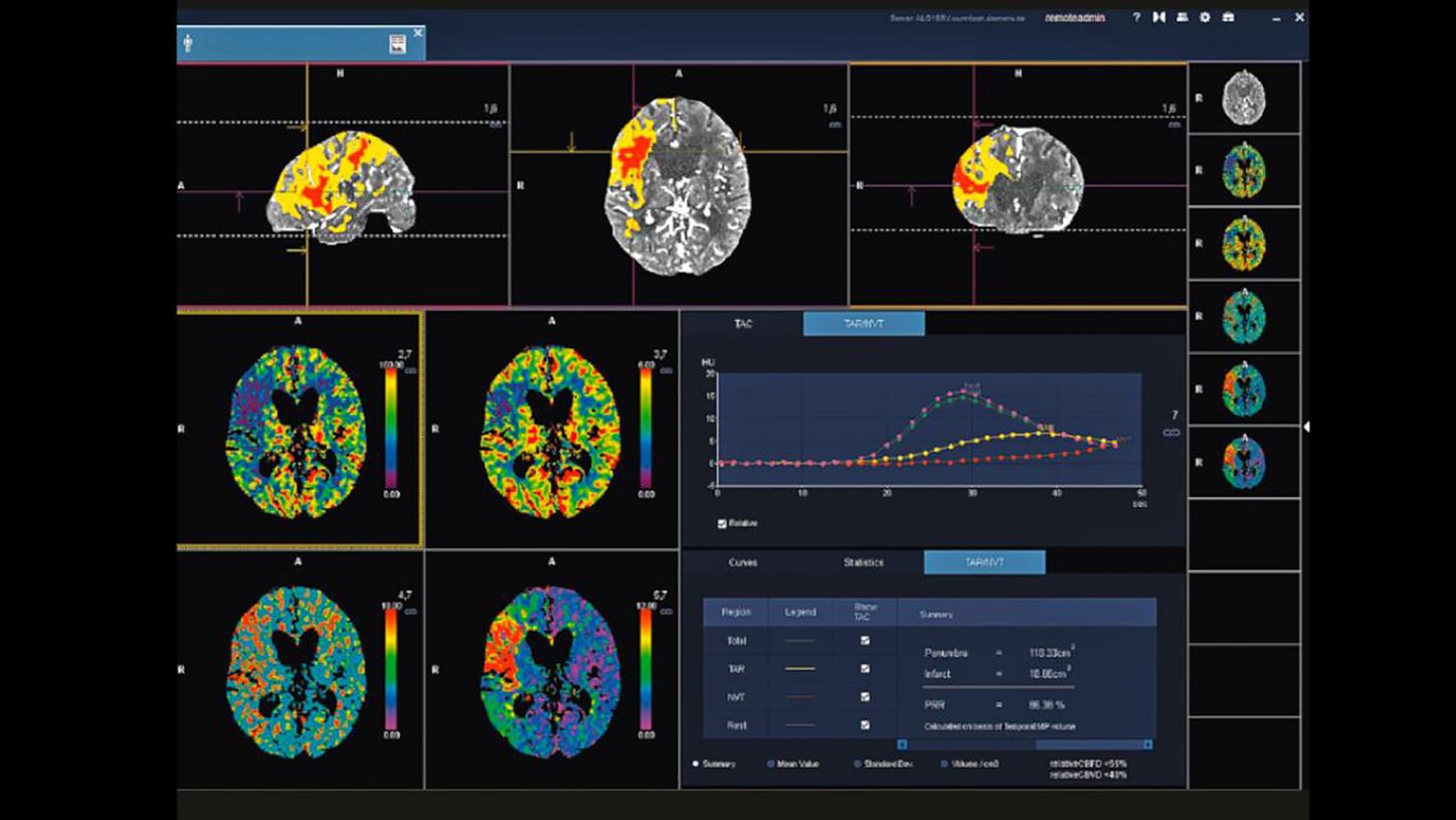
It takes just five simple steps to see the core infarct and penumbra. The guided workflow in syngo.CT Neuro Perfusion facilitates a routine 24/7 operation. Tissue at risk can be easily visualized in 3D color maps, based on the mismatch between cerebral blood volume (CBV) and cerebral blood flow (CBF). Or feel free to define a custom mismatch based on parameters you select.
Absolute leadership in 4D imaging. Smart, flexible, and dose-efficient.
The Adaptive 4D Spiral or Flex 4D Spiral technology offers whole-brain coverage and more for contrast-enhanced dynamic scans. It provides coverage beyond the detector width. On fixed CT gantries, it smoothly moves the table back and forth. With sliding gantry systems (SOMATOM Edge Plus and SOMATOM Definition Edge), the CT gantry moves back and forth on rails while the table remains fixed. This smart approach delivers more flexibility in z-coverage, allowing for industry-leading z-coverage over both small and large ranges. And it is more dose-efficient compared to large detector approaches.
Optimized stroke protocol with Adaptive 4D Spiral or Flex 4D Spiral
Minimize eye exposure – and get full diagnostic information
Our optimized stroke protocol is designed for routine 24/7 stroke imaging. It covers the whole supratentorial brain with 8.5 cm and up to 11.5 cm coverage.1 This minimizes eye exposure and risk of cataract at greater lengths2 – yet it provides full diagnostic information even though cerebellar infarcts are rare (<1%).3

Extended coverage may benefit specific cases
In certain clinical cases, it may be justified to increase z-coverage – and so we are offering an industry-leading z-coverage allowing up to aortic arch to vertex dedicated 4D CT-Angiography. Analyzing this data with syngo.CT Dynamic Angio may give you new insights that help better understand the dynamics of blood flow in neurovascular disease.
Enhance your clinical capabilities
Intelligent Dual Energy applications support faster evaluation of neurovascular diseases.

- Perform bone removal with fewer motion artifacts at lower dose
syngo.CT DE Direct Angio performs bone removal more reliably because it suffers less from the motion artifacts sometimes seen with DSA-based algorithms. It also achieves this at a lower dose, because a non-contrast CT is not required.

- Discriminate post-interventional bleeding from transient iodine uptake
When performing neuro-interventional procedures like clot retrieval in stroke, serious complications such as post-interventional bleeding can occasionally occur. On a single-energy CT, this can look similar to transient iodine uptake and show no clinical symptoms. This is where Dual Energy CT can be very helpful: syngo.CT DE Brain Hemorrhage helps discriminate between the two, and is now available on our single-source CT scanners.

- Evaluate faster with bones removed
In neurovascular disease evaluation and interventional treatment planning, syngo.CT Neuro DSA (Digital Subtraction Angiography) helps you save time and effort. Thanks to fully automated bone removal, you’ll find your images ready for reading as soon as you open a case; and you can toggle between bone and vessel views. The CT Neuro Package’s new CT Neurovascular workflow also permits a comprehensive vessel analysis of the head and neck – including curved planar reformations (CPR) for stenosis measurement and automated vessel tracking.
