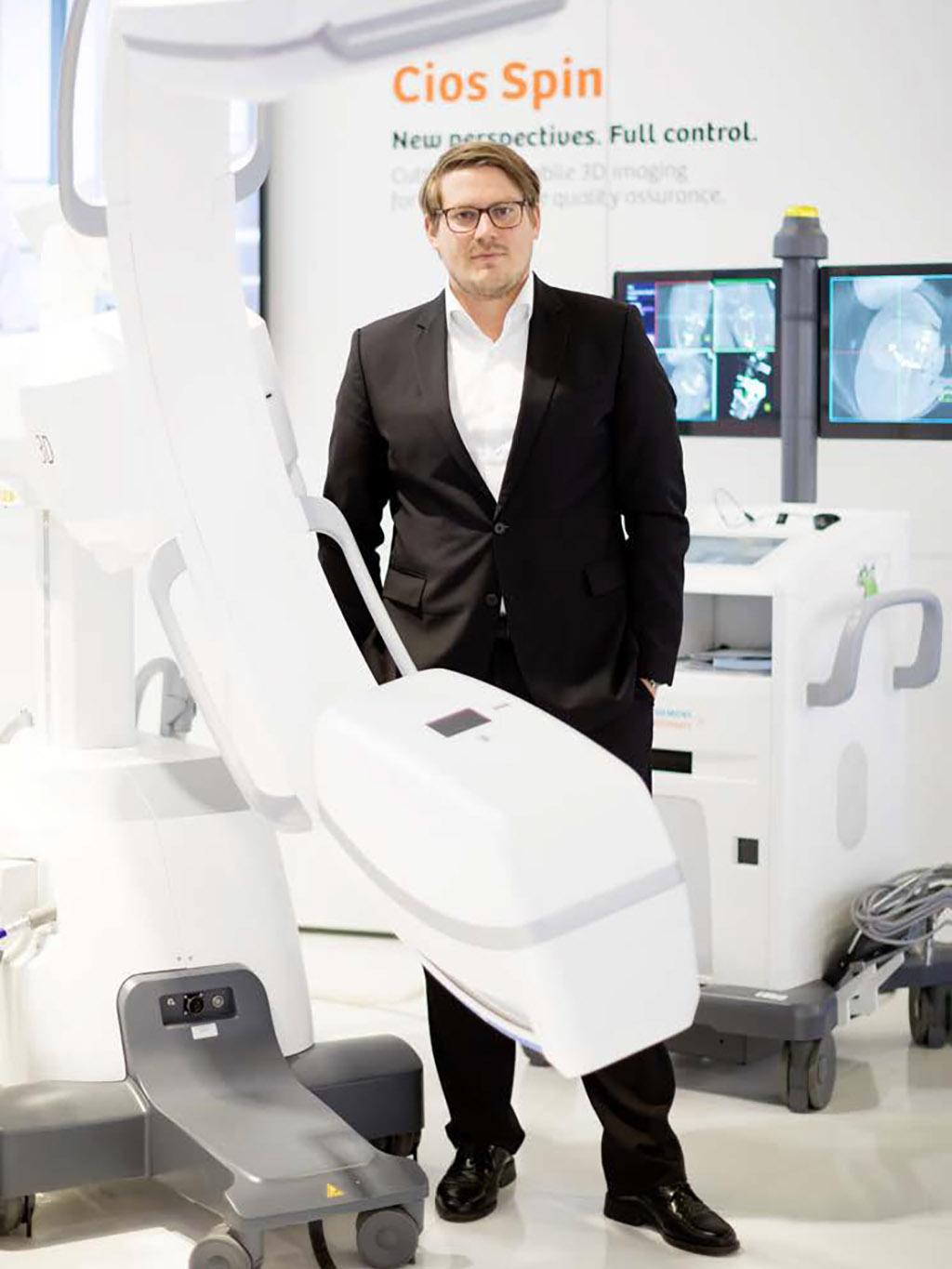
Intraoperative 3D imaging in orthopedic and trauma surgery helps position implants and screws more accurately and to optimally reposition bone fragments, while also reducing complications and costly revision surgery. Depending on their specific requirements, orthopedics/traumatology departments can perform 3D imaging with mobile C-arms or robotic-assisted systems.
Photos: Gordon Welters
Intraoperative imaging gives the surgeon an opportunity to correct the position of bone fragments or the placement of screws and other implants during an operation and hopefully to improve the result of the surgery,” says Jochen Franke, MD, Head of the Division of Trauma at the BG Trauma Center Ludwigshafen in Germany at a specialist symposium of the German Congress of Orthopedics and Traumatology in Berlin. His colleague Peter Richter, MD, assistant head of pelvis and spine surgery of the Department of Trauma Surgery at the University Hospital Ulm, Germany, agrees: “I can avoid additional surgical interventions that could cause severe complications, such as infections or damage to other structures. That‘s the key advantage for me.”

Better operation results and lower radiation doses
Implant or bone fragment misplacements can be detected much more reliably with intraoperative 3D imaging than with 2D fluoroscopy as found in most operating theaters, says Franke. He gives the example of a high fibula fracture with rupture of the syndesmosis, the ligaments connecting the tibia and fibula: “In one in four patients, the bone fragments are not correctly positioned the first time, and this is very often not visible in 2D.” In Ludwigshafen, the bone fragments in these patients are temporarily fixed in place with a Kirschner wire, and the repositioning result is then monitored using intraoperative 3D imaging. The screws are only inserted when the 3D images indicate that everything is anatomically correct. “This means that we do not have to check the result again later. Postoperative CT imaging is no longer required, and that can sometimes even reduce the radiation dose,” Franke says.
There are numerous other examples: patients with fractures of the spine or pelvis as well as patients with spinal tumors greatly benefit from intraoperative 3D imaging. For instance, it can be life threatening for patients with upper cervical spine injuries if a screw inserted in the vertebral body accidentally breaches the vertebral artery canal. “With a modern 3D C-arm I can see immediately if the screw has been placed correctly, and that lets me sleep better at night,” says Franke.

Extensive proof of benefit in clinical studies
In the Orthopedics and Trauma Surgery Department of the University Hospital Ulm, the spine and pelvis are the main areas of application for intraoperative 3D imaging with a robotic-assisted C-arm. But other areas benefit from the technology as well: “We use 3D for all ankle fractures and have recently also begun using it for patients who need a thermoablation because of osteoid osteomas in hard to reach places. The more we work with 3D imaging and the better the systems become, the more we find patients who benefit greatly from it,” says Richter.
A number of clinical studies and patient case series, in which German trauma surgeons were heavily involved, have shown just how significant the benefits are. In the field of acute traumatology, for example, the use of intraoperative 3D imaging in a wide range of fracture types allowed the intraoperative placement of bone fragments or implants to be optimized in around one in five patients.[1] In lower leg syndesmotic fractures, the rate was just under a third in a patient series published by Franke,[2] and it reached 40 percent in patients with heel bone fractures.[3]
Clear data also exists for pelvic and spine surgery. This shows that in fractures of the hip socket the mean fracture gap following surgery was significantly smaller when intraoperative 3D imaging was used.[4] “At the same time, radiation levels were lower with intraoperative 3D scanning than with postoperative CT,” says Richter. Another recent study looked at patients who had screws inserted in their vertebrae. 3D imaging revealed a significant misplacement in every tenth screw. In most of these patients this was corrected immediately, thus avoiding a second operation.[5]
Modern 3D systems offer considerable freedom and large 3D volume
From a technical perspective, there are various possibilities for intraoperative 3D imaging. While trauma surgeons at the BG Trauma Center Ludwigshafen primarily rely on mobile 3D C-arms, the University Hospital Ulm uses – in addition to several mobile 3D C-arms – a robotic-assisted floor-mounted C-arm that produces 3D images in CT-like quality.
Interdisciplinary use favors robotic-assisted imaging system
Whether or not it makes sense to use a robotic-assisted system in addition to mobile 3D C-arms in orthopedic/trauma surgery depends to a large extent on the individual application scenarios and also on the medical facility‘s operational priorities beyond orthopedic/trauma surgery, according to Franke and Richter.
“Our focus is on trauma and tumor surgery on the spine and pelvis. For this we need a large field of view and sometimes high radiation doses,” explains Richter. This is where the limitations of mobile C-arms become clear. If vascular imaging or 3D low contrast resolution is required, for example in tumor surgery, an angiography system is the better choice.
At the University Hospital Ulm, not only orthopedic/trauma surgeons, but also vascular surgeons and neurosurgeons need a powerful 3D system. On average, the orthopedic/trauma surgeons now use the system two days a week, the vascular surgeons use it on a further two days and the neurosurgeons on one day. “The fact that we can also use it for vascular interventions was an important factor in our decision to acquire an angiography system. It allowed us to spread the costs, and it also means the operating room is used more,” Richter says.
Mobile 3D C-arms score thanks to their high flexibilit
Most mobile C-arms only provide contrast medium images of blood vessels in 2D, but they are very flexible: “A mobile C-arm can simply be wheeled over if a colleague in the next OR needs it. There are far fewer space problems with these systems, which is a great advantage,” says Richter. That is one of the reasons why Ulm‘s orthopedic trauma surgeons have several mobile 3D C-arms in use in addition to the robotic-assisted system: “At large medical facilities like ours the two systems complement each other very well. When it comes to simple ankle fractures, the mobile 2D/3D C-arm is perfectly adequate. But for pelvic operations where we need more power we turn to the robotic system,” says Richter.
In Ludwigshafen the situation is different. No vascular procedures are performed, and in most cases, 3D imaging is done with a mobile C-arm. If that proves inadequate, an intraoperative CT is used. However, thanks to modern systems this is becoming increasingly rare, according to Franke: “In many cases the mobile 3D C-arm achieves an image quality equivalent to that of a simple CT, so that even in spinal or pelvic surgery we can often do without intraoperative CT. The C-arm is also excellent for examining obese patients.”
Modern product solutions for intraoperative 3D imaging
Cios spin®
- Latest generation of mobile 3D C-arm
- Very large 3D volume of 16×16×16 cm
- Comfortable clearance of more than 93 cm between detector and radiation source
ARTIS pheno®
- One of the most advanced fixed robotic-assisted angiography systems on the market
- Generous space of 95.5 cm for surgical instruments between generator and detector
- 3D volume covering up to ten vertebrae
- Extremely fast with scan times of around five seconds in almost all locations
About the Author
Philipp Grätzel von Grätz is a Berlin-based freelance journalist with a focus on health and technology. He writes in German and English for various healthcare and technology publications in Europe and the US.

