syngo DualVolume


Enhance your decision making during interventions
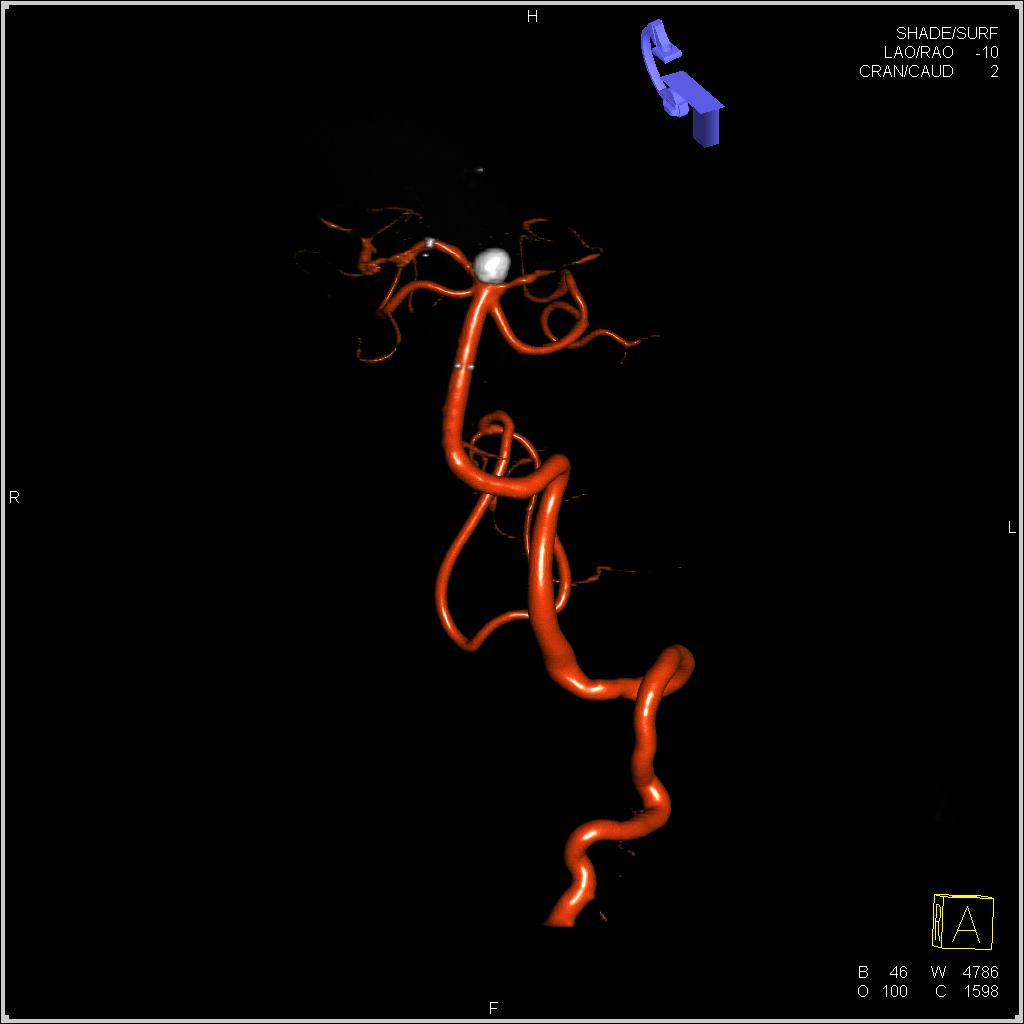
syngo® DualVolume enables the differentiation between two high-contrast 3D objects that have virtually the same contrast density. It supports clear visualization of a clip, stent, or coil placed in a vessel as well as soft restenosis within a stent and differentiates calcified plaque from a vessel.
Features & Benefits
syngo DualVolume allows the display of syngo DynaCT and 3D Angio in one view. Soft tissue information and anatomical landmarks are combined with contrast filled vessels. Anatomical structures of tumours can be seen in combination with its feeding vessels. This way syngo DualVolume provides a baseline for planning the embolization or surgical treatment of tumors.
Key Benefits
- Clearly differentiate between contrast-filled vessels, bones, stents and coils
- Evaluate stent deployment in complicated vessel pathologies
- Clearly see the anatomical structure of tumors in combination with the feeding vessels
- Identify complicated atypical vessel structures
- Visualize calcification next to contrast filled vessels
General Requirements
System
- Artis zee
- ARTIS pheno
Minimum Software Version
syngo X Workplace: VA60C
syngo MMWP: VD30B
Other
Please note: On account of certain regional limitations of sales rights and service availability, we cannot guarantee that all products included in this website are available through the Siemens sales organization worldwide. Availability and packaging may vary by country and are subject to change without prior notice. Some/All of the features and products described herein may not be available in all countries.
