- Home
- Medical Imaging
- Computed Tomography
- SOMATOM
- SOMATOM Edge Plus

SOMATOM Edge PlusChanging views in Computed Tomography
For care providers, the pressure to cut expenses results in an ever-increasing need for standardization and precision medicine. The CT scanner SOMATOM Edge Plus provides diagnostic imaging at the appropriate dose and with reproducible accuracy. Obtain rich, detailed information with technologies that bring tin-filtered scanning, 4D, and quantitative imaging to your clinical routine. And game-changing workflow automation simplifies scan preparation and helps you achieve new levels of precision.
Features & Benefits

Changing views on patient diversity – with personalized scanning
Scan virtually all patients with diagnostic confidence – including obese persons, children, and patients unable to cooperate.
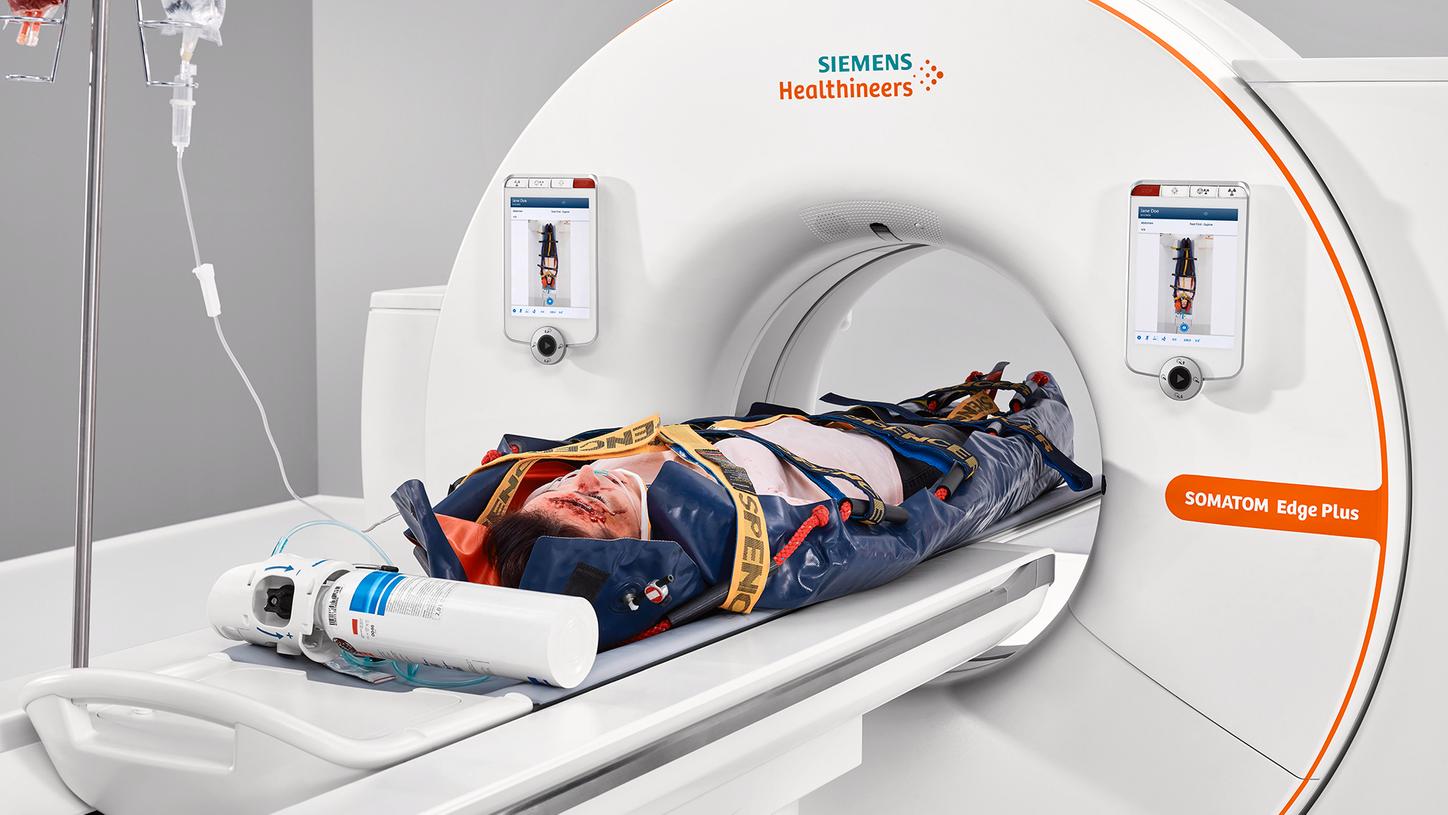


- Get powerful images of obese patients: With SOMATOM Edge Plus, you can obtain sharp and rich-in-contrast images at high speed and low dose even with large patients.
- Gently scan the dose-sensitive ones: Minimize the need for sedation and allow low-dose scans for children with High Power at low kV and integrated CARE Child technology.
- Freeze motion when your patient can't: Excellent image quality within seconds for emergency cases due to the combination of High Power reserves with high coverage of up to 230 mm/s in clinical routine.
Changing views on clinical paradigms – with advanced imaging
Gain new diagnostic insights like functional information and tissue characterization, acquired with no dose or time penalty.
TwinBeam Dual Energy allows to perform advanced quantitative imaging with no dose penalty


TwinBeam Dual Energy allows to perform advanced quantitative imaging with no dose penalty


TwinBeam Dual Energy allows to perform advanced quantitative imaging with no dose penalty
- See more than ever before with tin-filtered scanning: Shield patients from clinically irrelevant dose and use CT for a wider application range with our Tin Filter technology.
- Add dynamic imaging to your clinical routine: Make dynamic imaging an everyday method for nearly all patients and anatomical regions at the right dose and contrast-to-noise ratio.
- Perform advanced quantitative imaging with no dose penalty: Eliminate additional non-contrast scans by creating virtual ones – backed by our unique TwinBeam Dual Energy technology.
Changing views on patient positioning – with automated workflows
Save time and achieve consistent results with smart automation for fast, precise positioning, scanning, and postprocessing.

FAST Integrated Workflow supports correct and consistent positioning


FAST Integrated Workflow supports correct and consistent positioning


- Safeguard correct and consistent positioning: Our FAST Integrated Workflow with FAST 3D Camera helps your team acquire first-time right scans and manage tight schedules.
- Get closer to your patients: Set parameters without leaving your patients and offer them individual care with our Touch Panels on eye-level.
- Simplify preparation of patients and scanner: Accelerate workflows and lower dose, helping you optimize process efficiency and improve patient outcome with our software packages.
Clinical Use
Neurological Imaging
03Thorax Abdomen Pelvic Imaging
04Cardiovascular Imaging
05Pediatric Imaging
01Musculoskeletal Imaging
02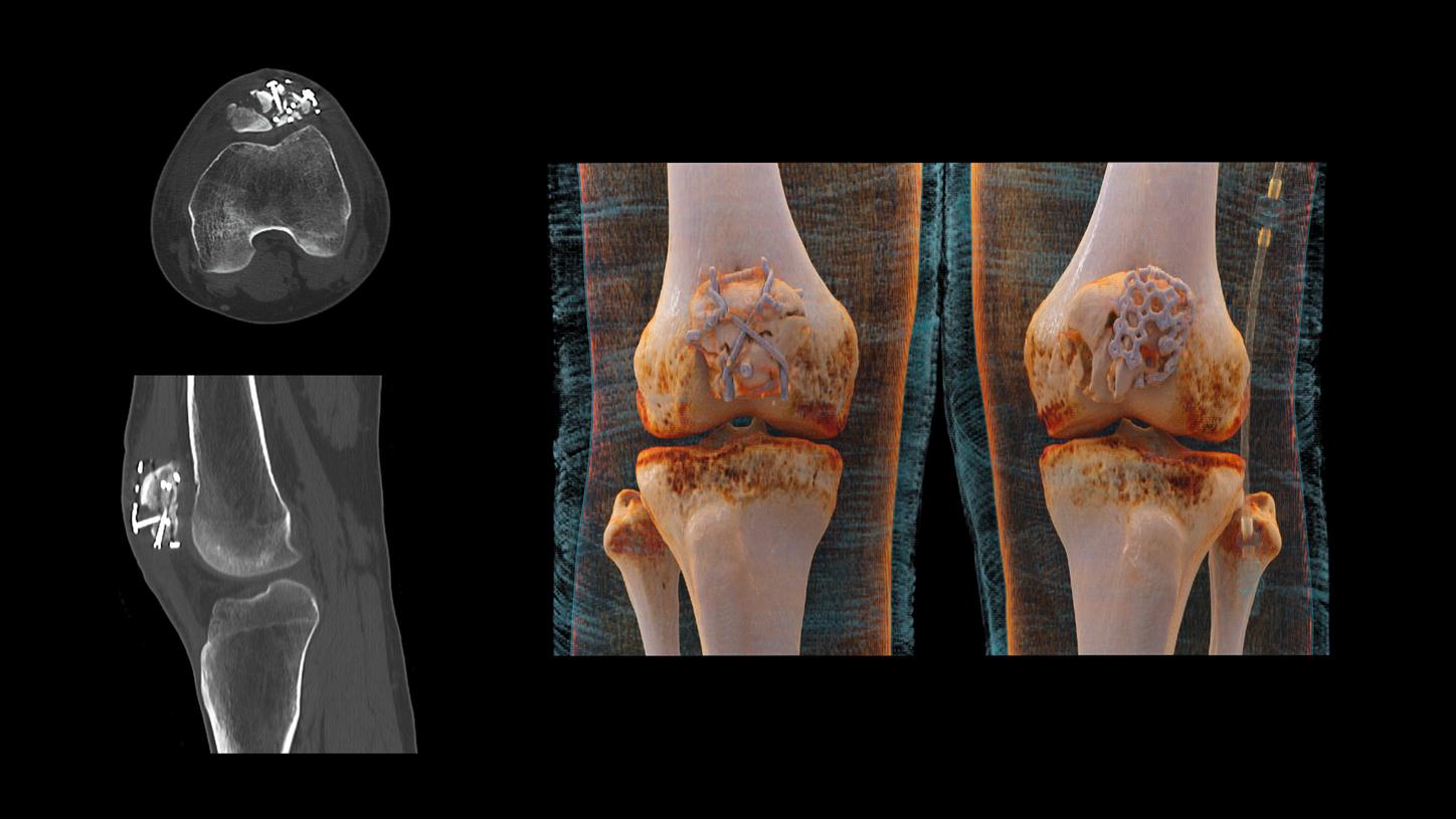
Sn140 kV
Scan time 10.35 s
Scan length 238 mm
CTDIvol 19.31 mGy
DLP 419.09 mGy*cm
- Native follow-up of fractured knees in cast
- Tin Filter for improved beam hardening
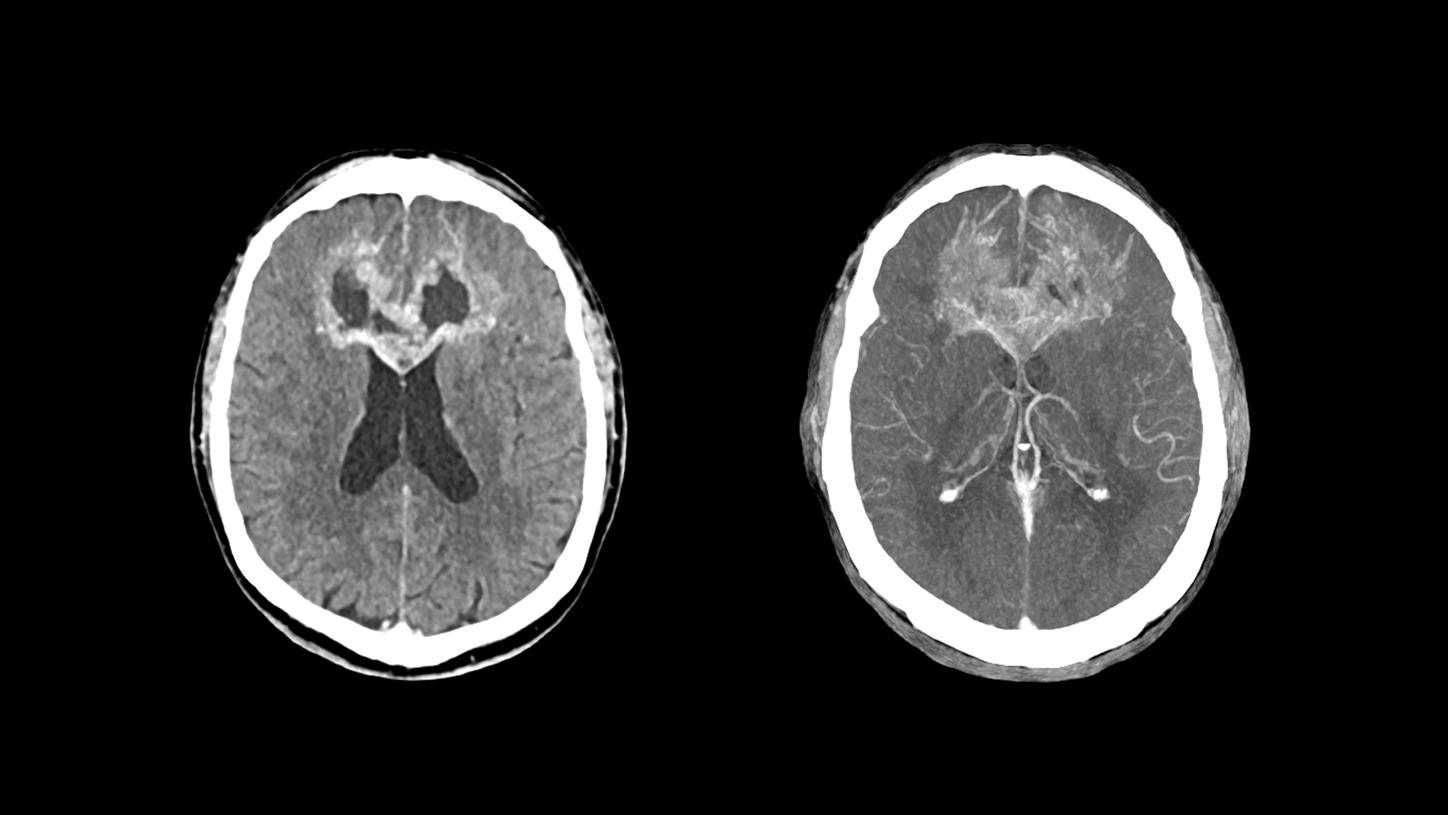
Courtesy of Erasmus MC, Rotterdam, The Netherlands
100 kV
Scan time 10.05 s
Scan length 212 mm
CTDIvol 38.05 mGy
DLP 731.68 mGy*cm
- Contrast media-enhanced scan of the brain for tumor staging and therapy planning
- Excellent image quality thanks to StellarInfinity detector

Sn100 kV
Scan time 6.16 s
Scan length 130 mm
CTDIvol 4.62 mGy
DLP 50.88 mGy*cm
Eff. Dose 0.1 mSv
- High image quality at very low dose levels can be achieved using an Sn100 kV (Tin Filter) protocol with iterative reconstruction
- Effective dose comparable to that of conventional radiography1

CTA
100 kV
Scan time 6 s
Scan length 389 mm
CTDIvol 9.4 mGy
DLP 339 mGy*cm
Perfusion
70 kV
Scan time 44 s
Scan length 118 mm
CTDIvol 151 mGy
DLP 1,780 mGy*cm
- Follow-up after clipping
- Adaptive 4D Spiral enables full brain perfusion imaging at low kV
- High Power at low kV enables high contrast-to-noise ratio and potential dose savings
- StellarInfinity detector enables high-resolution imaging of vascular regions
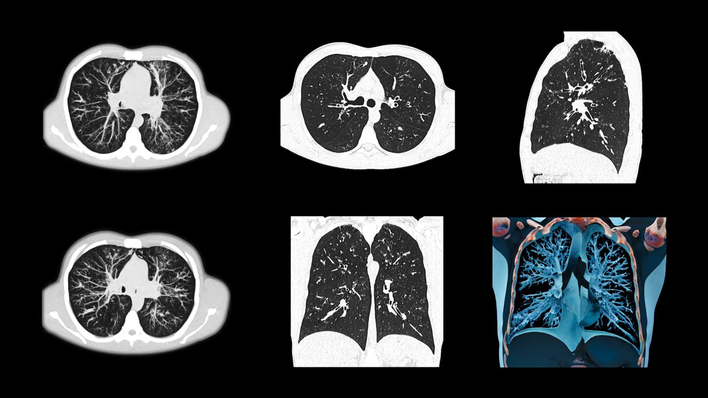
Sn100 kV / Sn100 kV
Scan time 7.73 s / 3.03 s
Scan length 356 mm / 300 mm
CTDIvol 1.65 mGy / 0.38 mGy
DLP 54.98 mGy*cm / 10.47 mGy*cm
- Tin-filtered low-dose imaging of the lung for evaluation of infiltrates
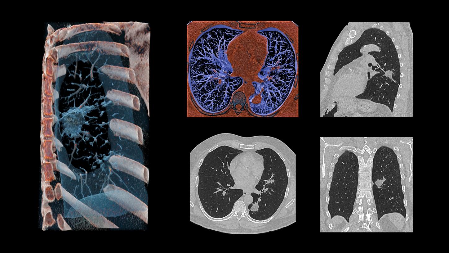
110 kV
Scan time 5 s
Scan length 474 mm
CTDIvol 9.43 mGy
DLP 414 mGy*cm
- Native lung imaging for lesion measurement and evaluation
- 10 kV Steps and High Power enable high contrast-to-noise ratio with potential dose savings

100 kV
Scan time 11 s
Scan length 661 mm
CTDIvol 12.9 mGy
DLP 819 mGy*cm
- High Power at low kV enables high contrast-to-noise ratio and potential dose savings
- Wide bore size enables scanning of even obese patients
- HD field of view enables visualization of tissue even out to 78 cm
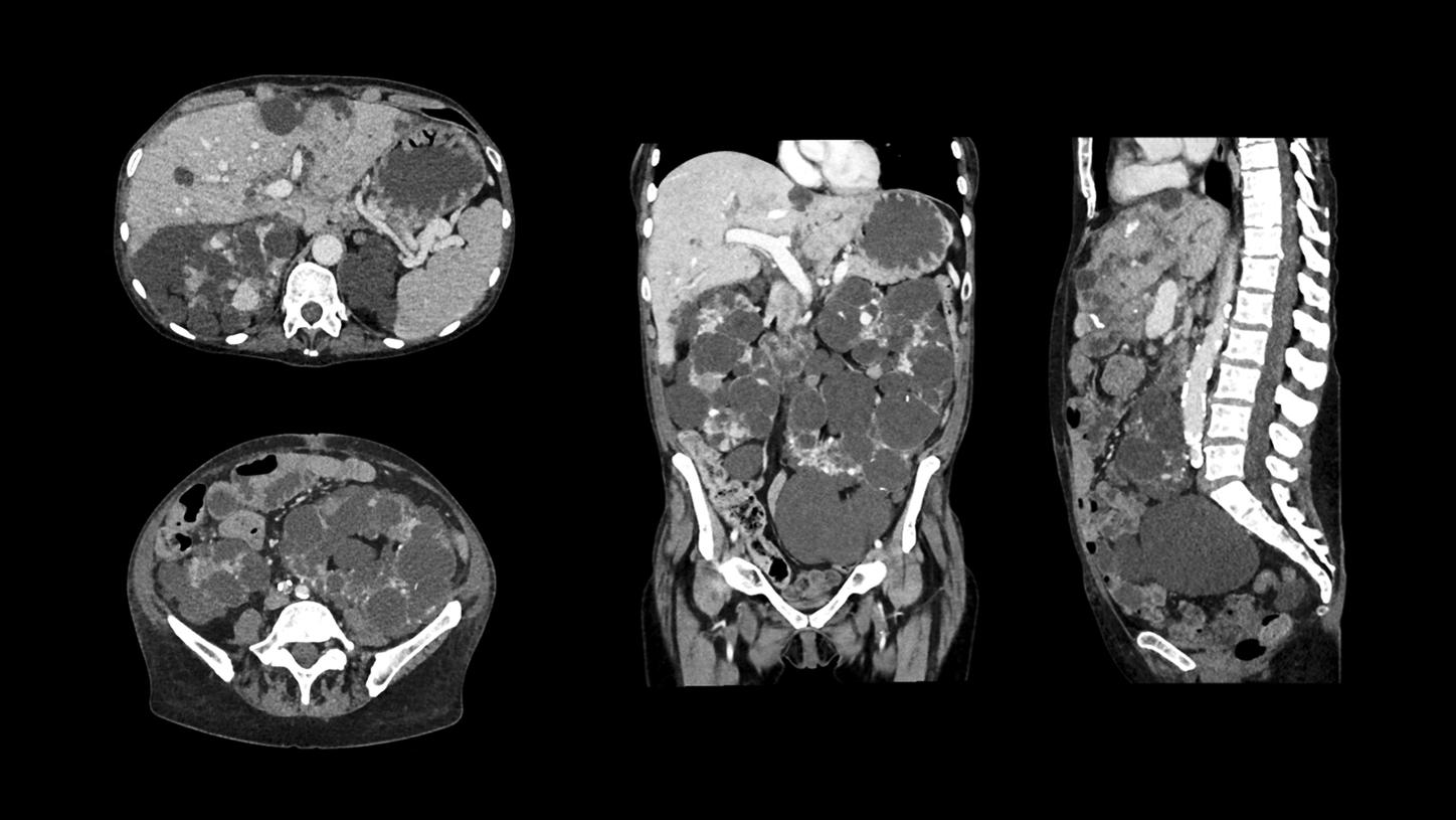
70 kV
Scan time 15.54 s
Scan length 535 mm
CTDIvol 8.15 mGy
DLP 421.56 mGy*cm
- Single-phase contrast media-enhanced scan of the abdomen and pelvis for evaluation of multi-cystic disease
- Great contrast-to-noise ratio at low dose due to High Power 70
- 1 mm slices shown
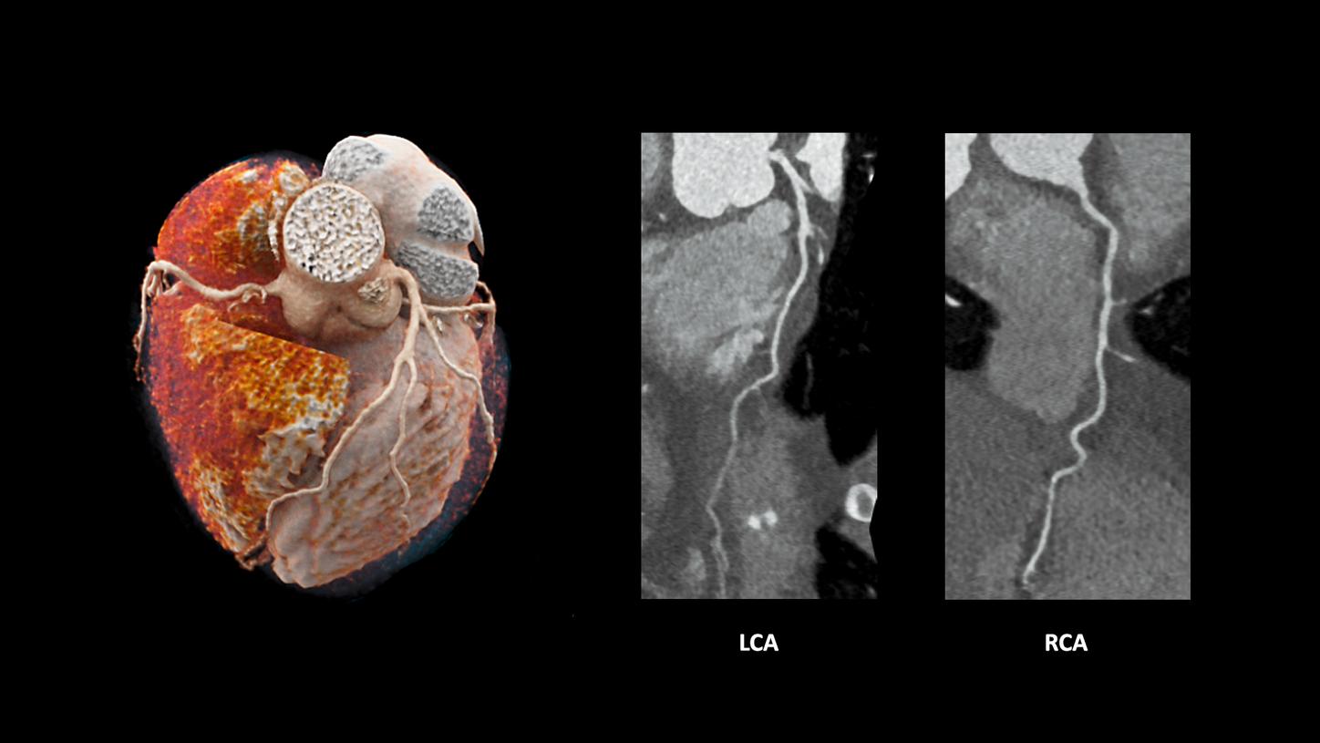
80 kV
Scan time 1.24 s (seq. acq.)
Scan length 104 mm
CTDIvol 5.32 mGy
DLP 56 mGy*cm
HR 55 bmp (avg.)
- Coronary CTA of patient with stable heart rate to rule out stenosis
- High Power at low kV enables high contrast-to-noise ratio and potential dose savings
- Improve intra-operator image quality reliability with FAST Phase
80 kV
Scan length 240 mm
CTDIvol 35.8 mGy
DLP 1,035 mGy*cm
- 4D CTA for follow-up of endovascular repair of an abdominal aortic aneurysm
- High Power at low kV enables high contrast-to-noise ratio and potential dose savings
- 4D scan range not limited by detector width
- Increase diagnostic certainty with 4D information

70 kV
Scan time 3 s
Scan length 677 mm
CTDIvol 1.45 mGy
DLP 94 mGy*cm
- Whole-body CTA of the aorta for assessment of aortic aneurysm
- High Power at 70 kV enables high contrast-to-noise ratio images with potential radiation and contrast media dose savings1
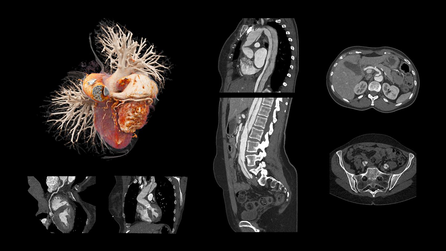
ECG-triggered thorax CTA
80kV
Scan time 1.96 s
Scan length 242 mm
CTDIvol 2 mGy
DLP 48.19 mGy*cm
Abdomen CTA
100 kV
Scan time 2.59 s
Scan length 454 mm
CTDIvol 4.81 mGy
DLP 203.58 mGy*cm
- Low-kV two-step aortic CTA with ECG triggering for thoracic aorta
- High Power at low kV enables high contrast-to-noise ratio and potential dose savings
- Fast pitch of 1.7, up to 230 mm/s in clinical routine
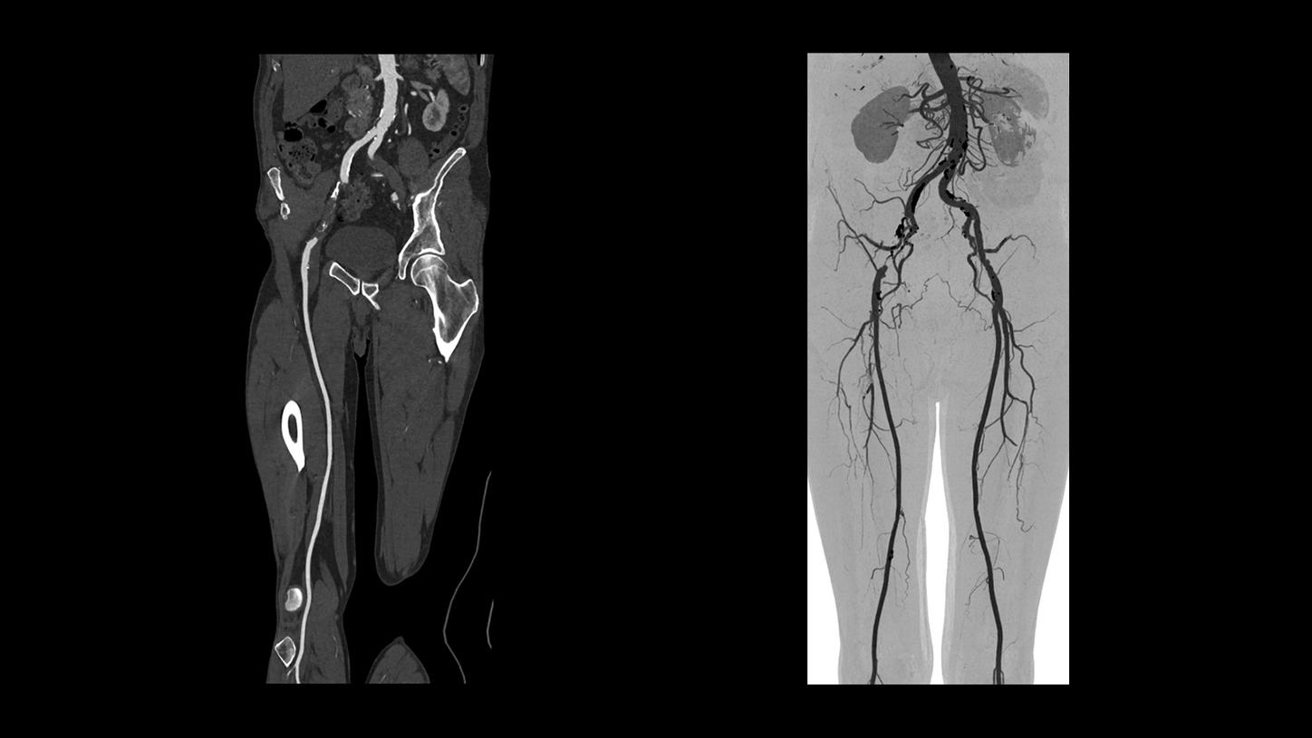
70 kV
Scan time 25.61 s
Scan length 1,349 mm
CTDIvol 1.84 mGy
DLP 243.69 mGy*cm
- Contrast media-enhanced CTA of the lower extremities for evaluation of arterial vessel disease
- High Power at 70 kV enabling high contrast-to-noise ratio images with potential dose savings
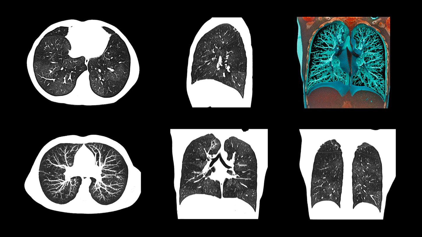
- Sn100 kV
- Scan time 2 s
- Scan length 314 mms
- CTDIvol 0.45 mGy
- DLP 12.7 mGy*cm
- Pediatric ultra-low-dose native lung imaging to assess infiltrates
- Tin Filter for lung evaluation at low dose

120 kV
Scan time 37.89 s
Scan length 171 mm
CTDIvol 34.4 mGy
DLP 582.43 mGy*cm
- Ultra-high-resolution scan of the knee after trauma with z-UHR comb

Sn140 kV
Scan time 10.35 s
Scan length 238 mm
CTDIvol 19.31 mGy
DLP 419.09 mGy*cm
- Native follow-up of fractured knees in cast
- Tin Filter for improved beam hardening

Courtesy of Erasmus MC, Rotterdam, The Netherlands
100 kV
Scan time 10.05 s
Scan length 212 mm
CTDIvol 38.05 mGy
DLP 731.68 mGy*cm
- Contrast media-enhanced scan of the brain for tumor staging and therapy planning
- Excellent image quality thanks to StellarInfinity detector

Sn100 kV
Scan time 6.16 s
Scan length 130 mm
CTDIvol 4.62 mGy
DLP 50.88 mGy*cm
Eff. Dose 0.1 mSv
- High image quality at very low dose levels can be achieved using an Sn100 kV (Tin Filter) protocol with iterative reconstruction
- Effective dose comparable to that of conventional radiography1

CTA
100 kV
Scan time 6 s
Scan length 389 mm
CTDIvol 9.4 mGy
DLP 339 mGy*cm
Perfusion
70 kV
Scan time 44 s
Scan length 118 mm
CTDIvol 151 mGy
DLP 1,780 mGy*cm
- Follow-up after clipping
- Adaptive 4D Spiral enables full brain perfusion imaging at low kV
- High Power at low kV enables high contrast-to-noise ratio and potential dose savings
- StellarInfinity detector enables high-resolution imaging of vascular regions

Sn100 kV / Sn100 kV
Scan time 7.73 s / 3.03 s
Scan length 356 mm / 300 mm
CTDIvol 1.65 mGy / 0.38 mGy
DLP 54.98 mGy*cm / 10.47 mGy*cm
- Tin-filtered low-dose imaging of the lung for evaluation of infiltrates

110 kV
Scan time 5 s
Scan length 474 mm
CTDIvol 9.43 mGy
DLP 414 mGy*cm
- Native lung imaging for lesion measurement and evaluation
- 10 kV Steps and High Power enable high contrast-to-noise ratio with potential dose savings

100 kV
Scan time 11 s
Scan length 661 mm
CTDIvol 12.9 mGy
DLP 819 mGy*cm
- High Power at low kV enables high contrast-to-noise ratio and potential dose savings
- Wide bore size enables scanning of even obese patients
- HD field of view enables visualization of tissue even out to 78 cm

70 kV
Scan time 15.54 s
Scan length 535 mm
CTDIvol 8.15 mGy
DLP 421.56 mGy*cm
- Single-phase contrast media-enhanced scan of the abdomen and pelvis for evaluation of multi-cystic disease
- Great contrast-to-noise ratio at low dose due to High Power 70
- 1 mm slices shown

80 kV
Scan time 1.24 s (seq. acq.)
Scan length 104 mm
CTDIvol 5.32 mGy
DLP 56 mGy*cm
HR 55 bmp (avg.)
- Coronary CTA of patient with stable heart rate to rule out stenosis
- High Power at low kV enables high contrast-to-noise ratio and potential dose savings
- Improve intra-operator image quality reliability with FAST Phase
80 kV
Scan length 240 mm
CTDIvol 35.8 mGy
DLP 1,035 mGy*cm
- 4D CTA for follow-up of endovascular repair of an abdominal aortic aneurysm
- High Power at low kV enables high contrast-to-noise ratio and potential dose savings
- 4D scan range not limited by detector width
- Increase diagnostic certainty with 4D information

70 kV
Scan time 3 s
Scan length 677 mm
CTDIvol 1.45 mGy
DLP 94 mGy*cm
- Whole-body CTA of the aorta for assessment of aortic aneurysm
- High Power at 70 kV enables high contrast-to-noise ratio images with potential radiation and contrast media dose savings1

ECG-triggered thorax CTA
80kV
Scan time 1.96 s
Scan length 242 mm
CTDIvol 2 mGy
DLP 48.19 mGy*cm
Abdomen CTA
100 kV
Scan time 2.59 s
Scan length 454 mm
CTDIvol 4.81 mGy
DLP 203.58 mGy*cm
- Low-kV two-step aortic CTA with ECG triggering for thoracic aorta
- High Power at low kV enables high contrast-to-noise ratio and potential dose savings
- Fast pitch of 1.7, up to 230 mm/s in clinical routine

70 kV
Scan time 25.61 s
Scan length 1,349 mm
CTDIvol 1.84 mGy
DLP 243.69 mGy*cm
- Contrast media-enhanced CTA of the lower extremities for evaluation of arterial vessel disease
- High Power at 70 kV enabling high contrast-to-noise ratio images with potential dose savings

- Sn100 kV
- Scan time 2 s
- Scan length 314 mms
- CTDIvol 0.45 mGy
- DLP 12.7 mGy*cm
- Pediatric ultra-low-dose native lung imaging to assess infiltrates
- Tin Filter for lung evaluation at low dose

120 kV
Scan time 37.89 s
Scan length 171 mm
CTDIvol 34.4 mGy
DLP 582.43 mGy*cm
- Ultra-high-resolution scan of the knee after trauma with z-UHR comb

Sn140 kV
Scan time 10.35 s
Scan length 238 mm
CTDIvol 19.31 mGy
DLP 419.09 mGy*cm
- Native follow-up of fractured knees in cast
- Tin Filter for improved beam hardening


Are you interested in more clinical cases? We created a booklet that contains a collection of different cases and clinical images – all acquired with SOMATOM Edge Plus.
Technical Specifications
Detector: | StellarInfinity detector |
Number of acquired slices: | 128 |
Number of reconstructed slices: | 384 |
Spatial resolution: | 0.30 mm |
Rotation time: | 0.28 s |
In-plane temporal resolution: | 142 ms |
Generator power: | 100 kW |
kV steps: | 70, 80, 90, 100, 110, 120, 130, 140 kV |
Max. scan speed: | 23 cm/s |
Table load: | up to 307 kg / 676 lbs |
Gantry opening: | 78 cm |

Digital education with PEPconnect
Accelerate you or your staff’s workflow and knowledge with PEPconnect and PEPconnection*. Engage in learning activities at any time and on any device for a personalized learning experience with PEPconnect. Using a PEPconnection subscription, you can access a workforce education management plan as well as analytics and progress report tracking.
*Subscription required. Availability of subscription depends on country.
Did this information help you?
Thank you.
Lell, et.al. Optimizing Contrast Media Injection Protocols in State-of-the Art Computed Tomographic Angiography. Investigational Radiology 2015; Volume 50, Number 3, March 2015.

