- Home
- Medical Imaging
- Computed Tomography
- CT Technologies & Innovations
- The Facts about Photon-counting CT

The Facts about Photon-counting CTGet to know the technology and the benefits
The introduction of photon-counting CT marks the beginning of a new era of precision medicine and clinical decision-making, thanks to a radically new approach to CT technology. For the first time, photon-counting CT directly converts X-rays into electrical signals—our NAEOTOM Alpha offers an entirely new way of generating clinical results that overcomes the limitations previously accepted as unavoidable.
But how exactly does photon-counting work? What are the facts, and what’s just marketing hype? Find out for yourself.
FACT: Photon-counting is more than just spectral imaging
Computed tomography scans offer high-resolution images for detailed information about anatomical structure. Scans can be performed at a speed that can capture a moving organ to empower a confident diagnosis. More recently, spectral information now provides insight into composition and function.
With conventional CT technology, it is not possible to get all three—resolution, speed, and spectral information—at the same time. Users are forced to compromise between high spatial resolution or low dose, high speed acquisition or spectral information.
With photon-counting CT, you can get all these things together in one acquisition—no compromises. Use the slider on the images below to see the difference the world’s first photon-counting CT can make.


Clinical image courtesy of Medical University of South Carolina, Charleston, SC, USA
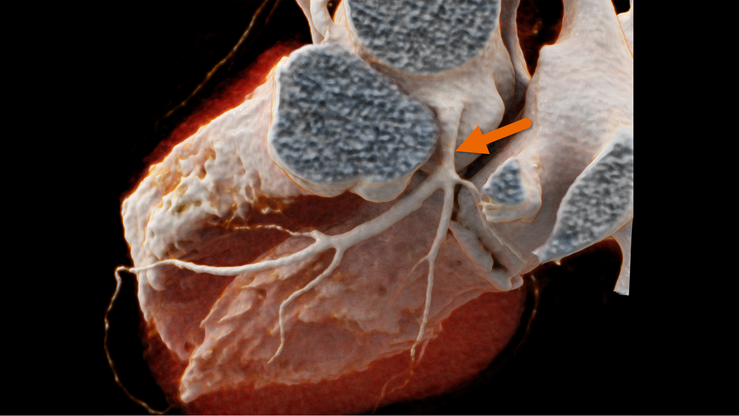
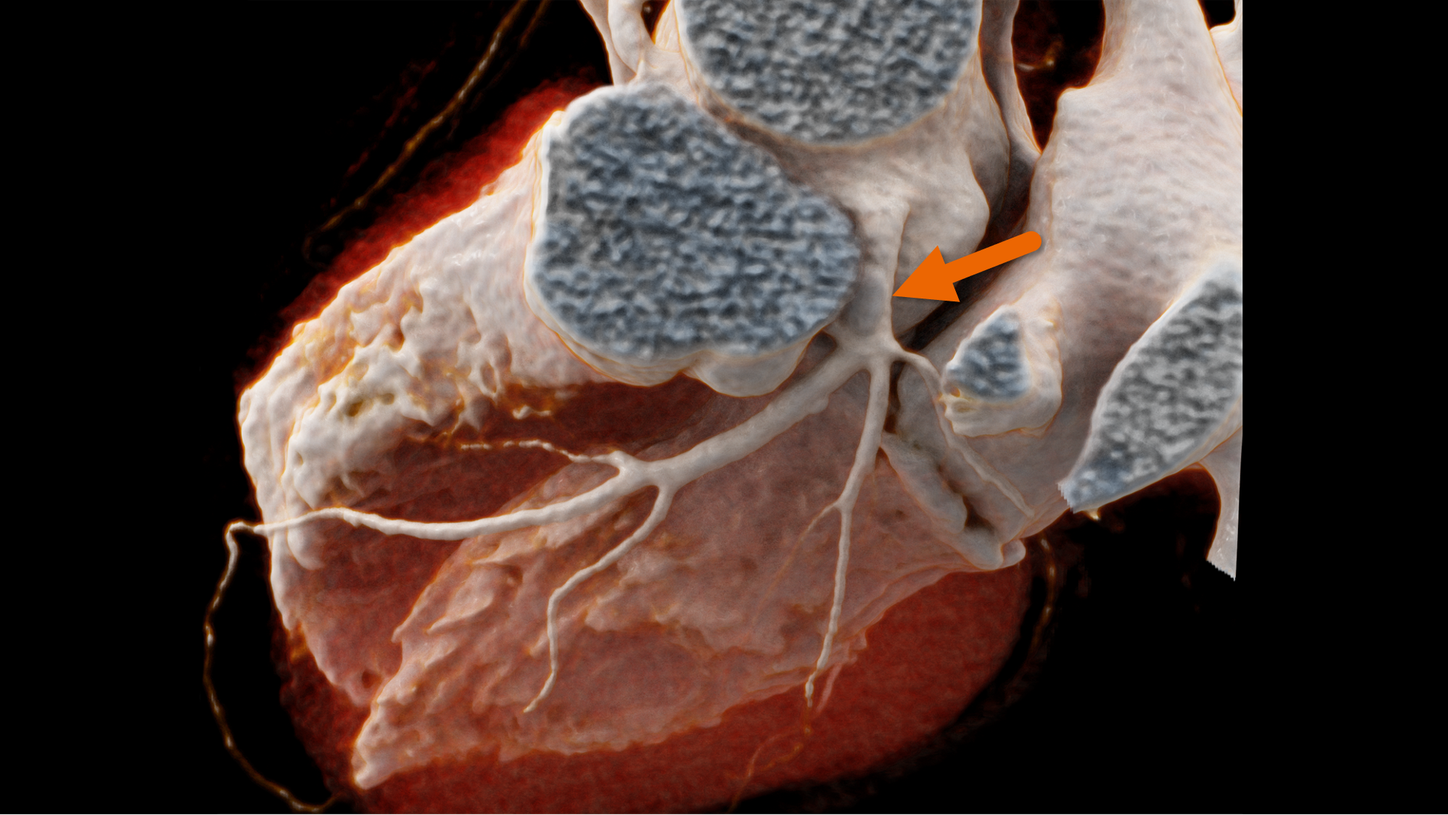
Scan patients previously excluded from cardiac CT
NAEOTOM Alpha offers impressive details for small structures and facilitates the differentiation of calcium from iodine.
 with
with without
Clinical image courtesy of Universitätsklinikum Augsburg, Augsburg, Germany
PURE Lumen
The artifacts resulting from calcifications in arteries can make the evaluation of the true lumen of a vessel impossible. PURE Lumen removes these calcifications based on the spectral information contained in every voxel of the image.
- Evaluate even heavily calcified vessels
- Create virtual non-calcium maps
- Perform coronary CTA exams also in patients with high CaScore


Clinical image courtesy of Erasmus Medical Center, Rotterdam, NL
Spectral imaging at high spatial resolution
With NAEOTOM Alpha, spectral maps are available more detailed than ever before—down to a slice thickness of 0.4 mm. This enables functional evaluation with high precision.
- Detect fine lesions in the liver
- Evaluate spectral bone marrow edema
- Evaluate lung perfusion defects based on detailed iodine maps


Clinical image courtesy of Medical University of South Carolina, Charleston, SC, USA
Scan patients previously excluded from cardiac CT
NAEOTOM Alpha offers impressive details for small structures and facilitates the differentiation of calcium from iodine.
 with
with without
Clinical image courtesy of Universitätsklinikum Augsburg, Augsburg, Germany
PURE Lumen
The artifacts resulting from calcifications in arteries can make the evaluation of the true lumen of a vessel impossible. PURE Lumen removes these calcifications based on the spectral information contained in every voxel of the image.
- Evaluate even heavily calcified vessels
- Create virtual non-calcium maps
- Perform coronary CTA exams also in patients with high CaScore


Clinical image courtesy of Erasmus Medical Center, Rotterdam, NL
Spectral imaging at high spatial resolution
With NAEOTOM Alpha, spectral maps are available more detailed than ever before—down to a slice thickness of 0.4 mm. This enables functional evaluation with high precision.
- Detect fine lesions in the liver
- Evaluate spectral bone marrow edema
- Evaluate lung perfusion defects based on detailed iodine maps


Clinical image courtesy of Medical University of South Carolina, Charleston, SC, USA
Scan patients previously excluded from cardiac CT
NAEOTOM Alpha offers impressive details for small structures and facilitates the differentiation of calcium from iodine.
 with
with without


Four fundamental advantages
Photon-counting CT provides intrinsic spectral sensitivity in every scan by measuring each X-ray photon against multiple energy thresholds. But that's just one piece of the puzzle. Explore more key advantages of photon-counting CT.
FACT: Photon-counting is a radically different detector technology
How does photon-counting work?
Every commercially available CT detector is based on the two-step principal of X-ray scintillation: X-ray to light, light to signal. The energy information derives from capturing multiple events simultaneously. The photo-counting detector, on the other hand, uses a crystal semiconductor instead of a ceramic scintillator. It directly generates an electric charge rather than creating light first. This one-step conversion from X-ray photon to electric signal enables an increased measurement speed. This means each of the 30,000 X-ray photons within a single projection can be measured individually.
More than dual layer
Dual layer detectors are not equivalent to photon-counting detectors. Dual layer detectors are still scintillator-based and require light as an intermediate step in converting X-rays to signals. They have the same physical limitations as all conventional energy integrating detectors.
In addition, dual layer detectors have the same challenge as single layer detectors when it comes to integrating electronic noise. The problem is amplified with a dual layer detector because of the interference with more layered components. Photon-counting, on the other hand, allows for the complete elimination of electronic noise.
FACT: NAEOTOM Alpha comes with simple, proven usability
The technology is complex. The workflow is not.
With such a dramatic change in the technology, one might expect photon-counting CT to add complexity and negatively impact your daily routine. However, through innovative technologies, including intelligent imaging guidance with myExam Companion and a mobile, tablet-based workflow, we have designed our photon-counting CT scanners to be simple and efficient to incorporate into broad clinical use.

Personalize procedures with tailored exam parameters and optimized dose

Standardize results with zero-click reconstructions
Empowering technologists with myExam Companion
myExam Companion guides operators through any CT scan procedure, so they can interact easily and naturally with both patient and technology. No matter the user, patient or throughput, it helps generate results that are both personalized and standardized.
myExam Companion is an intelligent imaging guidance that speaks your clinical language. It removes the guess-work and decision overload that frustrate users. It helps you quickly streamline the best exam options to generate high-quality consistent results for each patient.
Unleashing the full power of photon-counting
Our proven usability features support and guide users throughout the scanning workflow, while Al-enabled solutions help radiologists when reading cases.

Mobile Workflow with SOM X
Stay close to your patient with a tablet-based mobile workflow, with a simple, intuitive interface.

syngo.via
Unify and centralize intelligent tools in a powerful diagnostic workflow to make routine tasks convenient and efficient.

teamplay
A vendor-, system-, device-neutral digital health platform for fleet and protocol management in the cloud.
Learn more

High-Resolution Auto Mode
Balance the desire for details with the increased processing time required. Auto Mode selects the appropriate matrix size based on the FoV and Kernel.

GO Technologies
Standardize and simplify departmental processes from patient setup to image distribution, archiving, and reading with a holistic set of intuitive workflow solutions.

SPP Data Format
Always archive intrinsic spectral imaging source data, while minimizing the number of series sent to PACS thus reducing transfer time, image storage size and overall PACS clutter.
FACT: NAEOTOM Alpha is ready for primetime and making an impact
Abundant clinical evidence after a decades-long journey
Siemens Healthineers started basic research on photon-counting almost 20 years ago. The core of this technology is the semiconductor material—both the quality and the fabrication of the material. In 2012, based on their strong history and background in the production of CdTe crystals, we brought Acrorad into the Siemens Healthineers family.
The next step in the journey was working with our global collaboration partners to evaluate the technology from both technical and clinical perspectives. Through the installation of two generations of clinical prototypes beginning in 2014, these valued partners provided immeasurable insight into this groundbreaking technology. This journey culminated with the November 2021 launch of NAEOTOM Alpha: the world's first clinical photon-counting CT.
Innovation is at the core of Siemens Healthineers CT. Following the tradition of leading market innovations, including Dual Source CT, we are proud to be the first CT manufacturer to bring this technology to market.
We have now entered a new era of CT—and NAEOTOM Alpha is ready for primetime. Discover the full potential with these collections of photon-counting CT publications, symposia, and webinars.
User experience
Read what some of our early adopters have to say.
“This will redefine our clinical decision-making right from scan one.”1

Prof. Dr. med. Thomas Kröncke
University Hospital Augsburg, Germany
“There are so many applications on the horizon that will benefit our patients. I think it will make the medicine that we provide our patients more precise, and it will help us to provide truly patient-centered medicine.”1

Joseph Schoepf, MD
Medical University of South Carolina, Charleston, SC, USA
“A completely new way to make small structures of the heart visible.”1

Prof. Jiri Ferda
Charles University Pilsen, Czech Republic
“In the future, every CT will be a photon-counting CT.”1

Prof. Gabriel Krestin
Erasmus Medical Center Rotterdam, Netherlands
What photon-counting means for the future of your department
The NAEOTOM Alpha with Quantum Technology will redefine CT imaging. Based on the revolutionary direct signal-conversion of its QuantaMax detector, NAEOTOM Alpha offers high-resolution images at minimal dose, improved contrast at lower noise, and breakthrough consistency you’ve never seen before.

Scan previously excluded patients
The NAEOTOM Alpha may redefine which patient populations can be addressed with CT. It unlocks patient groups, like those with heavy calcifications, stents, pacemakers, or artificial valves, by offering intrinsic spectral imaging independent of scan speed, and of temporal or spatial resolution.
Impact treatment outcomes
Benefit from confident clinical decision-making in cardiology, oncology, and pulmonology, with answers that are meaningful, precise, and reproducible.
Did this information help you?
Thank you.
The statements by Siemens Healthineers’ customers described herein are based on results that were achieved in the customer's unique setting. Because there is no "typical" hospital or laboratory and many variables exist (e.g., hospital size, samples mix, case mix, level of IT and/or automation adoption) there can be no guarantee that other customers will achieve the same results.
