Artis Q is visionary in performance – with its high performance, advanced 3D imaging, and excellent contrast resolution at virtually any angle and for challenging patient sizes. And Artis Q is visionary in precision – supporting you in fighting the most threatening diseases such as coronary artery disease, stroke, and tumors. Expand precision medicine and take performance to the next level with Artis Q.
Artis QVisionary intervention
Artis Q is visionary in performance – with its high performance, advanced 3D imaging, and excellent contrast resolution at virtually any angle and for challenging patient sizes. And Artis Q is visionary in precision – supporting you in fighting the most threatening diseases such as coronary artery disease, stroke, and tumors. Expand precision medicine and take performance to the next level with Artis Q.
Features & Benefits
Visionary in performance
To see devices and anatomical structures even in challenging patients and angulations is one of the main goals in interventional imaging. Artis Q offers the technology you need for enhanced image quality, high contrast resolution even at steep angulations, and sharp images of moving objects – while reducing dose by up to 60% (GIGALIX compared to previous tube technology). Find out more about the technology inside Artis Q.

GIGALIX X-ray tube
Focused power
- Flat emitter technology for high contrast resolution even at steep angulations
- Small square focal spots for excellent spatial resolution to see more details
- CLEARpulse for sharp images and low dose

Large HDR detector
High dynamic range and dose efficiency
- High dynamic range for enhanced soft-tissue resolution in 3D imaging
- High dose efficiency enables excellent image quality at low radiation
- Water cooling to meet the demands of high hygienic standards
Clinical experience
Göttingen University Hospital, Germany
- Watch the movie to learn about Artis Freestyle transducer in clinical use
- Fast and easy sterile packing and highly flexible use
- Automatic patient data transfer from Artis Q to Artis Freestyle

“From the first procedures, we started to lower the fluoroscopy dose gradually and were surprised that we could actually go lower and lower – and the image quality was still good.”

Prof. Wacker
Head of Radiology, Medical School Hannover, Germany
Clinical Use
Visionary in precision
Precise guidance is needed to help improve clinical outcomes during interventions. Expand precision medicine with Artis Q – and find out more about the clinical applications and technologies it offers to support you in your clinical field.

Advanced 3D imaging – with syngo Dyna3D
- Differentiation between contrast-filledvessels and bones as well as devices byusing a mask and a fill run
- Dural arterial-venous fistula in 3D


Clinical image courtesy of University of Erlangen, Germany
1) Compared to previous software version VD11
Improved contrast and sharpness at the same dose level – with CLEAR MAX
- Improved contrast and sharpness lead to better visualization of details, small vessels, devices, tissue, and bones at the same dose level1)
- Separate processing of the device image leads to improved contrast and sharpness of devices in roadmap without affecting the vessel map1)
- Applicable to live imaging with DR, DSA, card acquisition, fluoroscopy, and roadmap

Clinical image courtesy of University Hospital Frankfurt, Germany
Evaluate perfusion for personalized therapy – with syngo DynaPBV Body
- Visualization of blood distribution directlyin the angio suite
- Supporting end point determination duringembolization
- Potential to identify nonresponders directlyafter interventional therapy

Clinical image courtesy of Hannover Medical School, Germany
Semi-automatic detection and targeted embolization – with syngo Embolization Guidance
- Automatic catheter and feeding-vesseldetection
- Easy one-click solution with tablesideoperation
- Vessel tree graphics overlay duringfluoroscopy for guidance

Clinical image courtesy of Hanover Medical School, Germany
Integrate the information of MRI, CT, or PET·CT into your angio image – with syngo Fusion Package
- Easy multi-modality integration withoutrequiring an intraprocedural 3D scan with syngo 3D/3D Fusion or syngo 2D/3D Fusion
- Overlay information from other modalitiesusing syngo 3D Roadmap or applicationslike syngo Toolbox with existing three-dimensional data sets

Clinical image courtesy of University Hospital Heidelberg, Germany
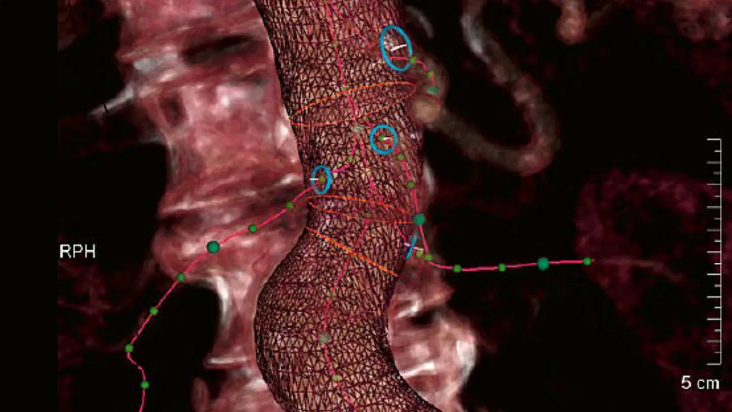
Automated workflow for endovascular repair – with syngo EVAR Guidance
- Automated detection of aorta wall and allbranched vessels
- Graphical representation of ostia rings andstent graft landing zones
- Automated image fusion and calculation ofoptimal C-arm angulations

Advanced 3D imaging – with syngo Dyna3D
- Differentiation between contrast-filledvessels and bones as well as devices byusing a mask and a fill run
- Dural arterial-venous fistula in 3D


Clinical image courtesy of University of Erlangen, Germany
1) Compared to previous software version VD11
Improved contrast and sharpness at the same dose level – with CLEAR MAX
- Improved contrast and sharpness lead to better visualization of details, small vessels, devices, tissue, and bones at the same dose level1)
- Separate processing of the device image leads to improved contrast and sharpness of devices in roadmap without affecting the vessel map1)
- Applicable to live imaging with DR, DSA, card acquisition, fluoroscopy, and roadmap

Clinical image courtesy of University Hospital Frankfurt, Germany
Evaluate perfusion for personalized therapy – with syngo DynaPBV Body
- Visualization of blood distribution directlyin the angio suite
- Supporting end point determination duringembolization
- Potential to identify nonresponders directlyafter interventional therapy

Clinical image courtesy of Hannover Medical School, Germany
Semi-automatic detection and targeted embolization – with syngo Embolization Guidance
- Automatic catheter and feeding-vesseldetection
- Easy one-click solution with tablesideoperation
- Vessel tree graphics overlay duringfluoroscopy for guidance

Clinical image courtesy of Hanover Medical School, Germany
Integrate the information of MRI, CT, or PET·CT into your angio image – with syngo Fusion Package
- Easy multi-modality integration withoutrequiring an intraprocedural 3D scan with syngo 3D/3D Fusion or syngo 2D/3D Fusion
- Overlay information from other modalitiesusing syngo 3D Roadmap or applicationslike syngo Toolbox with existing three-dimensional data sets

Clinical image courtesy of University Hospital Heidelberg, Germany
Automated workflow for endovascular repair – with syngo EVAR Guidance
- Automated detection of aorta wall and allbranched vessels
- Graphical representation of ostia rings andstent graft landing zones
- Automated image fusion and calculation ofoptimal C-arm angulations

Advanced 3D imaging – with syngo Dyna3D
- Differentiation between contrast-filledvessels and bones as well as devices byusing a mask and a fill run
- Dural arterial-venous fistula in 3D


Clinical image courtesy of University Hospital Erlangen, Germany
Boosting the level of detail – with syngo DynaCT Micro
- 40% increased spatial resolution compared to standard syngo DynaCT
- Better visualization of fine structures
- Enhanced evaluation of, e.g., stents, flow diverters, or stapes prosthesis

1)This is the experience of individual users. Results mayvary.
Welcome the 4th dimension to the angio suite –with syngo Dyna4D
- Based on this protocol a virtually unlimited number of DSA runs at no additional dose and contrast medium is provided.
- No additional contrast media compared to standard 3D1)
- Optimizing patient selection and individualized treatment strategies

Clinical image courtesy of University of Magdeburg, Magdeburg, Germany
Visualization of fine details – with GIGALIX X-ray tube
- Visibility of small vessels is increased by upto 70% compared to previous X-ray tubetechnology
- High contrast and spatial resolution forexcellent image quality
- Micro focal spot enables high detailresolution

Clinical image courtesy of University Hospital Magdeburg, Germany
Gray matter differentiation – with syngo DynaCT and 16-bit HDR detector technology
- Homogeneous image representation
- Excellent soft-tissue resolution
- Visualization of bleedings

Reduce metal artifacts to see the unseen – with syngo DynaCT SMART
- Reduced artifacts from dense objects
- Relevant aspects in soft tissue are madevisible even if they are close to, e.g., coilpackages or glue

Confident coil placement – with Advanced Roadmap
- Separate windowing for vessel and devicecontrast
- Select any DSA reference image as aroadmap
- Observe the glue injection or actual coilplacement with the vessel map from theinitial roadmap using the ‘Show Progress’function

Clinical image courtesy of University Hospital Erlangen, Germany
Boosting the level of detail – with syngo DynaCT Micro
- 40% increased spatial resolution compared to standard syngo DynaCT
- Better visualization of fine structures
- Enhanced evaluation of, e.g., stents, flow diverters, or stapes prosthesis

1)This is the experience of individual users. Results mayvary.
Welcome the 4th dimension to the angio suite –with syngo Dyna4D
- Based on this protocol a virtually unlimited number of DSA runs at no additional dose and contrast medium is provided.
- No additional contrast media compared to standard 3D1)
- Optimizing patient selection and individualized treatment strategies

Clinical image courtesy of University of Magdeburg, Magdeburg, Germany
Visualization of fine details – with GIGALIX X-ray tube
- Visibility of small vessels is increased by upto 70% compared to previous X-ray tubetechnology
- High contrast and spatial resolution forexcellent image quality
- Micro focal spot enables high detailresolution

Clinical image courtesy of University Hospital Magdeburg, Germany
Gray matter differentiation – with syngo DynaCT and 16-bit HDR detector technology
- Homogeneous image representation
- Excellent soft-tissue resolution
- Visualization of bleedings

Reduce metal artifacts to see the unseen – with syngo DynaCT SMART
- Reduced artifacts from dense objects
- Relevant aspects in soft tissue are madevisible even if they are close to, e.g., coilpackages or glue

Confident coil placement – with Advanced Roadmap
- Separate windowing for vessel and devicecontrast
- Select any DSA reference image as aroadmap
- Observe the glue injection or actual coilplacement with the vessel map from theinitial roadmap using the ‘Show Progress’function

Clinical image courtesy of University Hospital Erlangen, Germany
Boosting the level of detail – with syngo DynaCT Micro
- 40% increased spatial resolution compared to standard syngo DynaCT
- Better visualization of fine structures
- Enhanced evaluation of, e.g., stents, flow diverters, or stapes prosthesis


Minimize patient dose – with GIGALIX X-ray tube
- Reduction of patient dose of up to 60% compared to previous tube technology
- Increase of prefiltration
- Excellent temporal resolution to enhance acquisition of moving organs

Clinical image courtesy of AZ Maria Middelares, Ghent, Belgium
Real-time stent enhancement – with CLEARstent Live
- Support of complex procedures
- Real-time verification of stent positioning while moving the device
- Potential to speed up procedures and to save contrast agent*

Clinical image courtesy of Oulu University Hospital, Finland
Valve positioning support with syngo Aortic Valve Guidance
- Automated aortic root segmentation and visualization of anatomical landmarks in seconds
- Automated C-arm positioning to orthogonal view without fluoroscopy
- Improved guidance through overlay ofaortic contour and landmarks onto live 2Dimage
Minimize patient dose – with GIGALIX X-ray tube
- Reduction of patient dose of up to 60% compared to previous tube technology
- Increase of prefiltration
- Excellent temporal resolution to enhance acquisition of moving organs

Clinical image courtesy of AZ Maria Middelares, Ghent, Belgium
Real-time stent enhancement – with CLEARstent Live
- Support of complex procedures
- Real-time verification of stent positioning while moving the device
- Potential to speed up procedures and to save contrast agent*

Clinical image courtesy of Oulu University Hospital, Finland
Valve positioning support with syngo Aortic Valve Guidance
- Automated aortic root segmentation and visualization of anatomical landmarks in seconds
- Automated C-arm positioning to orthogonal view without fluoroscopy
- Improved guidance through overlay ofaortic contour and landmarks onto live 2Dimage
Minimize patient dose – with GIGALIX X-ray tube
- Reduction of patient dose of up to 60% compared to previous tube technology
- Increase of prefiltration
- Excellent temporal resolution to enhance acquisition of moving organs


“With the Artis Q the low-contrast resolution has significantly increased and facilitates the detection of small bleedings.”

Johannes Weber, MD
Head of Department of Diagnostic and Interventional Neuroradiology, Kantonsspital St. Gallen, Switzerland
“The biggest advantage of the Artis Q is its ability to take patients from the CT to the angio lab and perform formerly CT guided procedures with syngo DynaCT and iGuide1).”

Gerard O’Sullivan, MD
Department of Radiology, University College Hospital Galway, Ireland
Technical Details
2021 edition of Artis Q
Visionary intervention – that’s what Artis Q stands for. With techniques and technologies constantly changing, clinical institutions need a flexible angiography system that can be easily adapted. As an investment for the future, Artis Q empowers you to perform both routine and complex procedures today and to be ready for tomorrow's new procedures.

Latest operating system Windows 10
- Windows 10 provides latest technology to safeguard against increasing cybersecurity threats
- Future virus protection updates ensured

High performance imaging system hardware
- Upgraded hardware enables new live image edge enhancement
- New hardware of Artis imaging system and syngo X Workplace (with syngo Application Software) match requirements of new operating system software

Improved contrast and sharpness with CLEAR MAX
- Improved contrast and sharpness lead to better visualization of details, small vessels, devices,tissue, and bones at the same dose level2)
- Separate processing of the device image leads to improved contrast and sharpness of devices inroadmap without affecting the vessel map2)
Avez-vous jugé cette information utile?
Merci beaucoup
The results and statements by Siemens Healthineers customers described herein were achieved in the customer's unique setting. Since there is no "typical" hospital and many variables exist (e.g. hospital size, case mix, level of IT adoption), there can be no guarantee that other customers will achieve the same results.
Compared to previous software version VD11
Cette page est réservée uniquement aux professionnels de santé. Si vous êtes un professionnel de santé, confirmez en cliquant sur OK.


