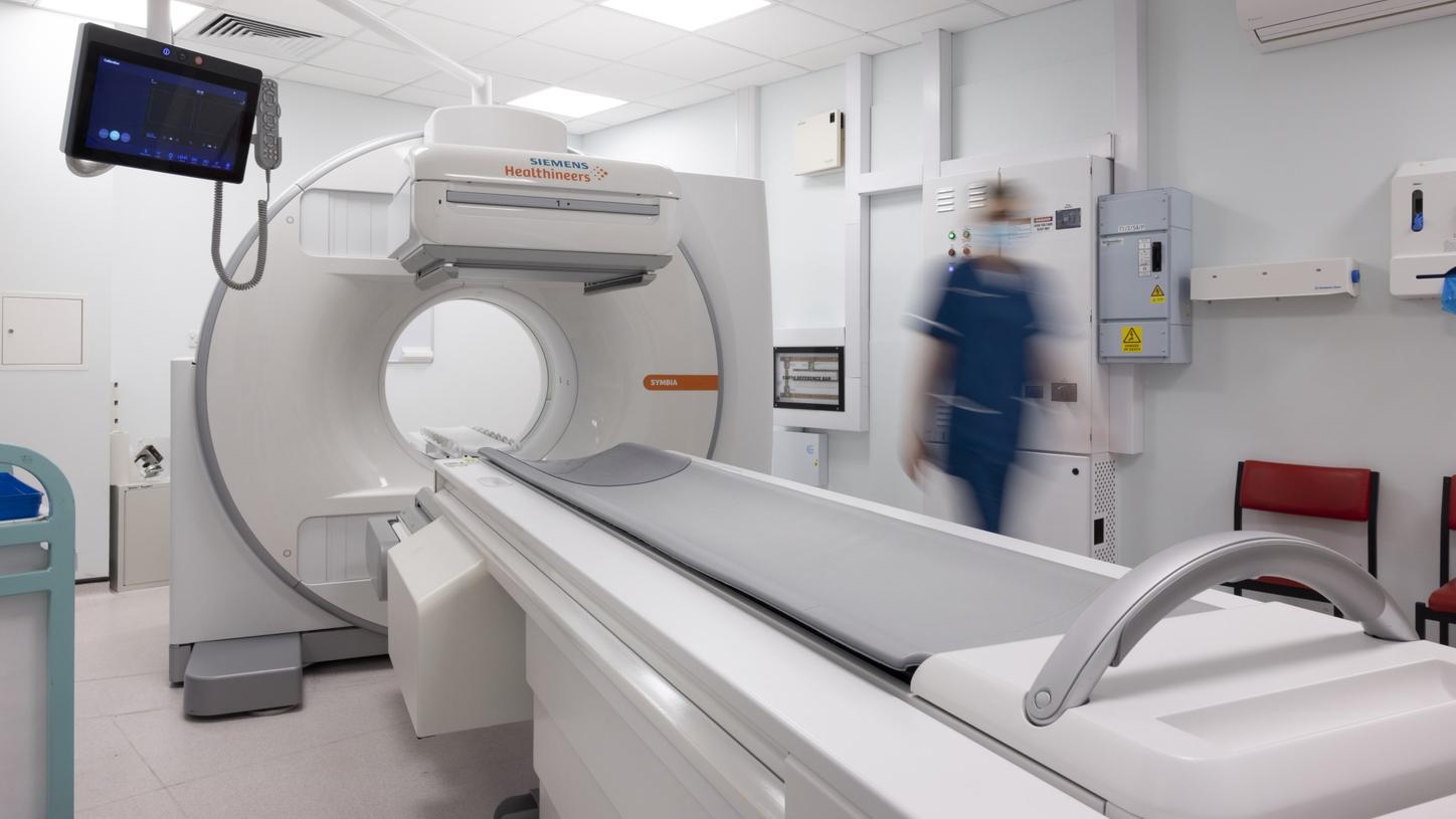
Nuclear medicine experts on two continents describe how the new fully integrated SPECT/CT serves more patients and signals the beginning of a new era in nuclear imaging.
Photography by Jonathan Browning and Scott van Osdol
Single-photon emission computed tomography (SPECT) combined with computed tomography (CT) was introduced as a hybrid SPECT/CT imaging modality almost two decades ago. Soon after the first commercially available diagnostic SPECT/CT system, Siemens’ Symbia TruePoint™ system, was launched in 2004, researchers recognized the potential for improving image quality and incorporating quantitative image acquisition and reconstruction.
Alexander Hans Vija, PhD, who has been working
in the field for more than 25 years
and is currently head of SPECT
research at Siemens Healthineers
Molecular Imaging in Hoffman
Estates, Illinois, USA, recalls that
collaborative research led to Symbia
Intevo™, which featured standardized
quantification and the high-resolution
xSPECT Bone™ reconstruction
algorithm. “But from here, we wanted
to have a more integrated solution
and ultimately enable many more
imaging departments to install a
SPECT/CT system.” This would allow
more patients to access a wider range
of services.
"I believe this system moves the bar to a level where everybody can enter the SPECT/CT world."
Transitioning from SPECT to SPECT/CT
Vija’s hopes for Symbia Pro.specta have been realized at Baylor Scott & White (BSW) Medical Center-Temple in central Texas (USA), a part of the BSW Health, one of the largest not-for-profit healthcare systems in the US. “We had been wanting a SPECT/CT camera for many years,” admits Michael L. Middleton, MD, director of the nuclear radiology division which employs three nuclear physicians and six technologists. Obtaining approval for purchase of a SPECT/CT system was challenging, however, due to reimbursement issues. “Approval of parathyroid SPECT/CT opened the door,” he recalls, but even then, installation of a SPECT/CT system was delayed for years. “We were left with just two Symbia™ SPECT-only cameras. We desperately needed a third nuclear medicine camera just to keep up with demand. So, Symbia Pro.specta was a godsend,” he says. The center has now been working with the new system for one year, and it has been providing more capabilities to a much wider variety of patients.
Expanded clinical applications
“The Symbia Pro.specta system has
been most useful for parathyroid
imaging, which we do once or twice
a day,” Middleton notes. “We also
schedule our larger (BMI >35) cardiac
patients on the camera because it
improves attenuation correction on
the myocardial perfusion scans in
these patients who have issues due
to soft tissue, large breasts, and fat.”
Women with breast cancer who have
had axillary breast surgery and cannot
raise their arms, causing attenuation, are also being moved to the Symbia
Pro.specta system. Additionally, it is
being used for octreotide scans where
patients do not have access to PET, for
MIBG scans, and imaging of 99mTc
MAA particles and bremsstrahlung
SPECT/CT for post-therapy imaging of
90Y microsphere selective internal
radiation therapy (SIRT) of liver
cancer. Another advantage of the new
camera and software is the improved
visualization of lesions compared with
planar imaging or SPECT alone, the
BSW professionals add. “With Symbia
Pro.specta, we have been able to start
using the xSPECT Bone reconstruction
algorithm, which helps us better
localize and characterize bone
disease,” Middleton says.
Another noticeable effect after upgrading to SPECT/CT was the intelligent imaging experience that fully integrates SPECT and CT. Middleton noticed how the staff appreciates the automation of the workflow. “It reduces the time going from the control room to the camera room. They can basically run it from the control room.” For lead nuclear medicine technologist Stephen Stoner, a big advantage of Symbia Pro.specta is streamlined design. “We don’t have all the big computers that we used to have. That gives us more space within the control room itself,” he says. “You just have the monitors and the keyboard on top of the table, with no big computer box underneath.”
“With Symbia Pro.specta, we have been able to start using the xSPECT Bone reconstruction algorithm, which helps us better localize and characterize bone disease.”
Middleton adds, “Any site acquiring one of these SPECT/CT cameras needs to schedule extra training time for the technologists to learn the new SPECT syngo® software platform. But then I think they will be happy with it.” Middleton’s team switched the software they used to read the scans to syngo.via. “It wasn’t a hard transition, and it’s been an improvement, especially on the cardiac reading,” he comments. “Overall, it’s been a positive learning curve.”
Upgrading for practiced SPECT/CT users
Across the Atlantic, in the United Kingdom, staff in the department of nuclear medicine at the Queen Elizabeth Hospital Birmingham (QEHB) in the UK Midlands have been using SPECT/CT for over 10 years. When the hospital opened as part of the University Hospitals Birmingham NHS Foundation Trust in 2010, the department had a Symbia S SPECT, Symbia T SPECT/CT, and a Symbia T16 SPECT/CT. The Symbia T16 has since been replaced and the hospital now has a range of SPECT/CT scanners. In February 2020, a Symbia Pro.specta system was installed, which occupies the space where formerly a Symbia S SPECT camera was installed.
Erin Ross, PhD, consultant clinical scientist and deputy head of nuclear medicine at QEHB, recalls that when the hospital first opened, the radiologists had an open mind about using SPECT/CT. “As soon as they started using it, they embraced it and they kept putting everything onto the Symbia T16 due to the higher quality of CT.” For many years since, Ross and her colleagues have been asking for “better CT, the best images in the quickest time, a diagnostic “one-stop shop” to overcome “a real blockage in efficiency,” she says. “Now we’ve got such a high quality of SPECT/CT with Symbia Pro.specta.” Khalid Hussain, MD, consultant radiologist at QEHB, confirms that “because of the high quality of the SPECT, we have been able to reduce the dose, which is excellent.” He adds that “the quality of the CT as part of the SPECT/CT is “much higher” than they have ever seen before.
A new SPECT/CT feature for the QEHB staff is iterative reconstruction on the CT. “The SAFIRE (sinogram affirmed iterative reconstruction) algorithm has also helped us lower doses in the CT examinations,” Ross says. “Tin Filter has also significantly lowered CT doses as compared to the other machines.” The technologists also appreciate the iMAR (iterative metal artifact reconstruction) technique, which they are using for the first time.
Providing a comfortable experience
is important for technologists who
engage with patients directly.
During patient transfer, patients are
less worried about falling off
because the bed is wider. In the
scanning room, the position of the
handset on the gantry display is
popular. Laura Whitehouse, senior
clinical technologist specializing in
nuclear medicine, appreciates that
the camera is smaller, quieter, and
less bulky when it’s moving around
the patient. “That’s really important
for patients who are nervous,” she
says. “They get a better experience,
so they’re more relaxed.” She also
praises the incorporation of the CT
component. “It’s all within the
casing, and we don’t have to bother with turning the CT on and off.
Everything’s more compact, clean,
smart, and user-friendly,” she says. ”I
love that I’m not kicking the
hardware box underneath the
control room desk any longer,”
Ross adds.
For principal clinical technologist
Yasmin Wahid, a welcome new
feature on Symbia Pro.specta is the
pre-recorded breathing instructions
in different languages for the
patients. Whitehouse also likes the
appearance of the new, one-color
interface, which she believes is
important for patients when they
first enter the room. “It’s really
appealing. It’s smaller, and it doesn’t
look so bulky and scary,” she
comments. For Wahid, it’s simpler
and easier, “It’s literally just one
button and we’re ready to go.”
Intelligent imaging experience at QEHB
Everyday operations in the exam
room are streamlined with smart
workflows. Whitehouse likes that the
workflows themselves are easier to
set up. “When you’re on the fly, you
can chop and change whatever you
want,” she notes. “So, if the patient
tells you they have pain elsewhere
and if they’ve already had their
injection, we can simply just add
another scan. With the old system,
we would have one set workflow,
and if you wanted to add extra
images, you would have to complete
that workflow and reload another.”
She adds that with the Plan&GO, an
intuitive bedside ruler for scan range
planning (“We call it ‘the magic
ruler’”), it is easier to identify
landmarks and position the patient.
As the lead of the nuclear cardiology
service, Wahid appreciates that the
gantry display (Scan&GO) is a new
touchscreen, so that it is easy to
focus the heart in the central field of
view instead of having to click
through a series of tabs to see
different positions. “If we want to
change the collimator, and we want
to see the patient’s ECG, now we
have everything on one screen,” she
enthuses. She also highlights the
reduction in time needed for cardiac
scans with Symbia Pro.specta,
especially for ECG-gated scans to
measure left ventricular function.
This is particularly useful for
monitoring cardiac side effects of
chemotherapy and greatly improves
the patient experience. “With some
patients, like those with an irregular
heartbeat (arrhythmia), it would take
us absolutely ages to acquire the data,” Wahid recalls. “Now we can do
a quicker scan, so instead of being on
the bed for about 35 minutes, it’s
now more like 15 minutes.”
“Now we’ve got such a high quality of SPECT/CT with Symbia Pro.specta.”
With Symbia Pro.specta and its smart workflow, QEHB nuclear medicine services can now be expanded, allowing greater numbers of SPECT/ CT scans to be fitted into their schedule. These include post-therapy SPECT/CT for detection and characterization of radioactive 131I focal uptake in patients with thyroid cancer. Octreotide scans have been restarted in support of the PET service. Significant expansion of SPECT/CT scans being done post 177Lu DOTATATE therapy for neuroendocrine tumors. Increased numbers of FLR (future liver remnant) scans are being done for preoperative assessment, having been moved from the Symbia T2. The SIRT service for colorectal cancer and hepatocellular carcinoma has also been moved from the T2, because of the higher quality of the CT with Symbia Pro.specta. Even for practiced SPECT/CT technicians, change can be challenging—but worth it. Whitehouse cautions that the Symbia Pro.specta interface is completely different from previous Symbia systems. “At first it can be a little daunting, but when you get used to it, everything is much more user-friendly,” she says.
Reaching more patients through expanded use of Symbia Pro.specta SPECT/CT
The UK and USA medical facilities are looking forward to expanding their services with Symbia Pro.specta. “Our dream would be to get up and running with contrast-enhanced CT, for follow-up prostate or bone scans, and be able to go one-stop shop with that. That is our aim for next year,” Ross says.
The positive experience of both the
US and UK hospitals with the new
Symbia Pro.specta reflects the
culmination of Vija’s vision of
wider availability and easier use,
but also demonstrates the potential
for both nuclear medicine
departments to expand their patient
services even beyond those they
envisaged when they first started
using the Symbia Pro.specta.
About the author
Linda Brookes is a freelance medical writer and editor. She divides her time between London and New York, working for a variety of clients in the healthcare and pharmaceutical fields.


