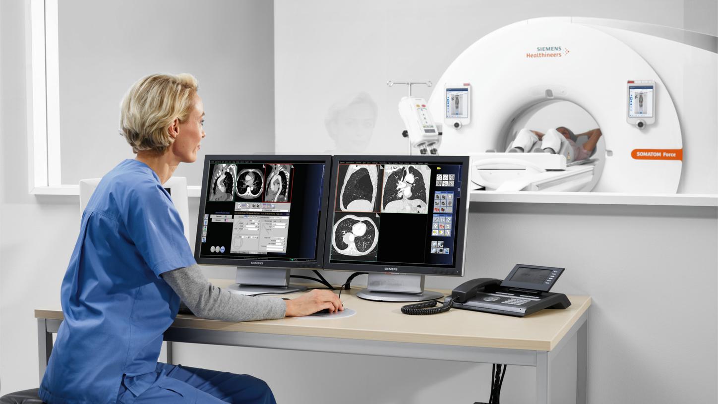
Nearly one third of deaths worldwide is caused by cardiovascular disease.1 Cardiovascular imaging is considered to be one of the central pillars of diagnostic imaging, and Computed Tomography plays its part here significantly. With the increase in referrals for these procedures comes an increase in the complexity of the patients needing scans. We offer perfectly attuned solutions to answer your clinical questions in Cardiology – from diagnosis to therapy and follow-up.
- Home
- Medical Imaging
- Computed Tomography
- Clinical Imaging Solutions
- CT imaging in cardiology

CT Cardiovascular imagingThe full spectrum of state-of-the art cardiac CT solutions
Features & Benefits
Redefine CT in Cardiology with NAEOTOM Alpha: Patient Reach. Unlocked.
NAEOTOM Alpha redefines which patient populations can be addressed with CT. It offers spectral imaging independent of scan speed and of temporal or spatial resolution.
This way, NAEOTOM Alpha makes it possible to confidently examine previously excluded patients, to perform scans that were not practicable before, and to increase follow-up frequency if desired. More clinical options. More opportunities for growth.
Explore NAEOTOM Alpha, the world’s first photon-counting CT, and other cardiac benefits beyond patient reach.

Ready beyond tomorrow with Dual Source CT
Clinical indications for cardiac CT are rapidly evolving. Already today it proceeded to much more than just coronary CTA: valvular disease, myocardial viability and perfusion, venous anatomy and congenital heart disease are just the beginning. This, along with patient diversity, requires the utmost flexibility in cardiac acquisition strategies – from systolic imaging through to full functional assessment at the lowest possible dose. The right acquisition for the right clinical indication and patient. Be ready beyond tomorrow with Dual Source CT.
Streamline your cardiac workflow with cardiac CT made easy
Cardiovascular CT imaging is often perceived as a challenging procedure. Optimal image quality is dependent on a combination of various factors such as patient preparation, ECG signal quality and optimal timing of scan in the most appropriate phase of the heart cycle. We make cardiac CT easy for you with our unique GO technologies combined with the unique mobile workflow.
Going to the Top of Cardiac CT imaging with SOMATOM go.Top
"Heart diseases remain the first cause of death in the western world. With cardiac CT we can assess coronary artery disease and according to the guidelines, now it's one of the first imaging modalities." Dr. Kallifatidis further states: "Our colleagues, our collaborators, cardiologists and cardiac surgeons, they are asking for more and more examinations. With our new scanner, the SOMATOM go.Top, we have completely changed our workflow and I have raised the amount of my work up to 30 to 40%."2
Simplify your routine with syngo.via and automatic post-processing
Pressure and workload in radiology are constantly rising: more cases per day, more cost pressure, higher expectations. As an intelligent, integrated imaging software syngo.via with the CT Cardiovascular applications offer the full spectrum of applications optimized for your clinical challenges. And applications combined with Rapid Results Technology it helps to account for workflow efficiency as well as standardization of quality of care by automatic image creation and filing.

Improve patient care – with High Power 70, 80 and 90 and ultra-low dose scanning
One of our persistent objectives is minimizing radiation exposure in cardiovascular imaging following the ALARA (As Low As Reasonable Achievable) principle. Additionally, the need for contrast media reduction e.g. for TAVI (transcatheter aortic valve intervention) scans of elderly and frail patients is increasing. Therefore, we want to equip you with the appropriate means such as our low kV capabilities or Tin Filter, e.g. for Calcium Scoring.


Identify significant coronary artery disease with CT based functional testing
Functional CT strategies offer additional incremental accuracy for detecting hemodynamically significant Coronary Artery Disease among patients with intermediate stenosis compared with anatomical assessment alone. This is achieved by the incremental improvement in specificity and positive predictive value afforded by e.g. dynamic stress CT perfusion , FFRCT (Fractional Flow Reserve) Analysis and Dual Energy perfusion.
Watch the video on the left to discover the benefits of dynamic myocardial perfusion.
Clinical Use

Pre-procedural evaluation of aortic stenosis for TAVI
Combination of high temporal resolution & low kV
SOMATOM go.Top
- Scan time: 4.5 / 4.7 s
- Scan length: 123 / 623 mm
- 80 kV
- CTDIvol: 6.02 / 1.88 mGy
- DLP: 74 / 118 mGy cm
- Clear visualization of aortic valve and coronary ostia in systole thanks to high temporal resolution
- Low dose scan of heart, aorta and vascular system with 80 kV

Dynamic myocardial stress perfusion to look for effect of a stenosis
SOMATOM Force
- Scan time: 33.41 s
- Scan length: 104.3 mm
- 70 kV, 275 mAs
- CTDIvol: 43.08 mGy
- DLP: 455.0 mGy cm
- Heart rate: 85-92 bpm
- 66 ms temporal resolution for systolic scanning potential
- Low kV imaging to keep dose as low as possible in 4D imaging

ECG triggered Turbo Flash cardiac scan with normal coronary arteries
SOMATOM Force
- Scan time: 0.12 s
- Scan length: 93 mm
- 80/80 kV, 289 mAs
- CTDIvol: 1.33 mGy
- DLP: 19.7 mGy cm
- Heart rate: 117 bpm
- Even with higher heart rates, such as here with 117 bpm, the Turbo Flash mode delivers high-quality clinical results with extremely low contrast media usage

2-month-old infant with heart anomaly at HR of 130 bpm
SOMATOM Force
- Scan time: 0.61 s
- Scan length: 79 mm
- 70/70 kV, 115 mAs
- CTDIvol: 1.16 mGy
- DLP: 9.1 mGy cm
- Heart rate: 130 bpm
- Vectron™ X-ray tube enables low kV scans even with high heart rates
- StellarInfinitiy detector reduces image noise and enhances spatial resolution

87-year-old patient for post stent follow-up
SOMATOM Drive
- Scan time: 4.4 s
- Scan length: 147 mm
- 80 kV
- CTDIvol: 18.91 mGy
- DLP: 277.1 mGy cm
- Eff. dose: 3.8 mSv
- Low kV study due to High Power 80
- High resolution stent imaging due to StellarInfinity detector

Courtesy of Erasmus MC, Rotterdam, The Netherlands
1This claim was supported based with the following CTA based study: Lell, et.al. Optimizing Contrast Media Injection Protocols in State-of-the Art Computed Tomographic Angiography. Investigational Radiology 2015; Volume 50, Number 3, March 2015.
Whole-body CTA of the aorta for assessment of aortic aneurysm
SOMATOM Edge Plus
- Scan time: 3 s each
- Scan length: 677 mm each
- 70 kV each
- CTDIvol: 1.45 mGy
- DLP: 94 mGy cm
- CARE Child and High Power enable high CNR images with potential radiation and contrast media dose savings1

Benefit of including HeartFlow FFRCT Analysis in workup of patients with suspected coronary artery disease
SOMATOM Drive
Patient history
- 73-year-old male
- Check-up due to chest pain on exertion
Scan findings
- Calcified and noncalcified plaque in all three vessels
- Moderate stenosis in LAD and LCX
Retrospectively analyzed with the HeartFlow FFRCT Analysis

Pre-procedural evaluation of aortic stenosis for TAVI
Combination of high temporal resolution & low kV
SOMATOM go.Top
- Scan time: 4.5 / 4.7 s
- Scan length: 123 / 623 mm
- 80 kV
- CTDIvol: 6.02 / 1.88 mGy
- DLP: 74 / 118 mGy cm
- Clear visualization of aortic valve and coronary ostia in systole thanks to high temporal resolution
- Low dose scan of heart, aorta and vascular system with 80 kV

Dynamic myocardial stress perfusion to look for effect of a stenosis
SOMATOM Force
- Scan time: 33.41 s
- Scan length: 104.3 mm
- 70 kV, 275 mAs
- CTDIvol: 43.08 mGy
- DLP: 455.0 mGy cm
- Heart rate: 85-92 bpm
- 66 ms temporal resolution for systolic scanning potential
- Low kV imaging to keep dose as low as possible in 4D imaging

ECG triggered Turbo Flash cardiac scan with normal coronary arteries
SOMATOM Force
- Scan time: 0.12 s
- Scan length: 93 mm
- 80/80 kV, 289 mAs
- CTDIvol: 1.33 mGy
- DLP: 19.7 mGy cm
- Heart rate: 117 bpm
- Even with higher heart rates, such as here with 117 bpm, the Turbo Flash mode delivers high-quality clinical results with extremely low contrast media usage

2-month-old infant with heart anomaly at HR of 130 bpm
SOMATOM Force
- Scan time: 0.61 s
- Scan length: 79 mm
- 70/70 kV, 115 mAs
- CTDIvol: 1.16 mGy
- DLP: 9.1 mGy cm
- Heart rate: 130 bpm
- Vectron™ X-ray tube enables low kV scans even with high heart rates
- StellarInfinitiy detector reduces image noise and enhances spatial resolution

87-year-old patient for post stent follow-up
SOMATOM Drive
- Scan time: 4.4 s
- Scan length: 147 mm
- 80 kV
- CTDIvol: 18.91 mGy
- DLP: 277.1 mGy cm
- Eff. dose: 3.8 mSv
- Low kV study due to High Power 80
- High resolution stent imaging due to StellarInfinity detector

Courtesy of Erasmus MC, Rotterdam, The Netherlands
1This claim was supported based with the following CTA based study: Lell, et.al. Optimizing Contrast Media Injection Protocols in State-of-the Art Computed Tomographic Angiography. Investigational Radiology 2015; Volume 50, Number 3, March 2015.
Whole-body CTA of the aorta for assessment of aortic aneurysm
SOMATOM Edge Plus
- Scan time: 3 s each
- Scan length: 677 mm each
- 70 kV each
- CTDIvol: 1.45 mGy
- DLP: 94 mGy cm
- CARE Child and High Power enable high CNR images with potential radiation and contrast media dose savings1

Benefit of including HeartFlow FFRCT Analysis in workup of patients with suspected coronary artery disease
SOMATOM Drive
Patient history
- 73-year-old male
- Check-up due to chest pain on exertion
Scan findings
- Calcified and noncalcified plaque in all three vessels
- Moderate stenosis in LAD and LCX
Retrospectively analyzed with the HeartFlow FFRCT Analysis

Pre-procedural evaluation of aortic stenosis for TAVI
Combination of high temporal resolution & low kV
SOMATOM go.Top
- Scan time: 4.5 / 4.7 s
- Scan length: 123 / 623 mm
- 80 kV
- CTDIvol: 6.02 / 1.88 mGy
- DLP: 74 / 118 mGy cm
- Clear visualization of aortic valve and coronary ostia in systole thanks to high temporal resolution
- Low dose scan of heart, aorta and vascular system with 80 kV


Did this information help you?
Thank you.
The statements by Siemens Healthineers’ customers described herein are based on results that were achieved in the customer's unique setting. Because there is no "typical" hospital or laboratory and many variables exist (e.g., hospital size, samples mix, case mix, level of IT and/or automation adoption) there can be no guarantee that other customers will achieve the same results.

