AI技術を用いて開発された最新の全自動撮影システム「myExam Companion」を搭載。個々の患者の状況に合わせた検査内容を自動で作成し、技師の経験に関わらず最適な検査の実施と一貫性のある結果を提供します。SOMATOM X. プラットフォームはハイパワーなX線出力が可能なVectron X線管によって、画質に妥協しない高速、低線量、高解像度のイメージングを可能にします。

myExam Companion 搭載
SOMATOM X. platformインテリジェントCT


一貫性を高めるインテリジェントなナビゲーション
ワークフローの自動化を支援する対話型アシスタント:限られた時間の中でも効率よく、オペレーターの経験に依存しない一貫したCT検査を実施できるようにサポートします。
myExam Companion
施設の運用に基づいて作成した質問事項に答えるだけで、検査準備から撮影、画像再構成、ポストプロセスに至るすべてのワークフローステップを最適化します。
myNeedle Companion
ニードルパスの計画とガイダンス、イメージフージョン、CTガントリ一体型のレーザーによって、CTガイド下インターベンションのワークフローを支援します。
myExam Companion
Intelligent CT workflow solution in action
myExam Companion が検査結果と自身のワークライフバランスに与える影響について、イタリアのナポリにあるセントロ・メディコ・アシオーネ・トッレ・デル・グレコのルチア・ラ・ムーラ博士の経験をご覧ください。
myNeedle Companion
ターゲットを絞ったニードルパス計画とガイダンス
米国アイオワ州デモインの放射線科でmyNeedle Companion による高度なレーザーガイダンスがCTガイド下インターベンションをどのように改善したかをご覧ください。

82cmのボア径を備えた患者に優しいデザイン
SOMATOM X. プラットフォームは、大容量の X-ray tube と耐荷重に優れる患者テーブル、82 cm のボア径を実現したことで、体格の大きな患者や、緊急検査に柔軟に対応することができます。高齢で検査体位に制限のある患者や、救急検査でフレキシブルなポジショニングが求められるケース、また、インターベンション手技においても患者や術者の特徴に応じて、安全にアプローチすることが可能です。
患者に優しいデザイン
システム設計により、さまざまな検査で患者さんがどのようにリラックスできるかをご覧ください。
ビジュアルエイド
検査を受ける全ての人のために、次世代のペイシェント・エクスペリエンスを実現します。

一貫性のある最適な結果をもたらすエリートパフォーマンス
SOMATOM X.プラットフォームは、難易度の高い検査でも、優れた空間・時間分解能と高速撮影、ポストプロセスの自動化技術を組み合わせることで、患者と疾患の両方の特性に合わせた個別化されたCT検査を実現します。Siemens Healthineers 独自のイメージングチェーンとユーザーガイダンスを融合することによって、優れたパフォーマンスを一貫して提供します。

SOMATOM X. プラットフォームによる高品質で低線量のイメージング

施設全体で一貫した操作性を実現
myExam Companion、myNeedle Companion、およびSiemens Healthineers のユーザーインターフェースであるShuiは、複数のモダリティにわたって共通の操作性を提供します。Siemens Healthinners のデジタルソリューションを組み合わせることで、施設全体のプロトコルを標準化し、ワークフローの改善と生産性の向上を実現します。
SOMATOM X. プラットフォーム


SOMATOM X.ceed
全ての検査でハイクオリティをスタンダードにする高解像度の高速CTです。
クリニカル情報ギャラリー
1/15
Cardiovascular Imaging03
Lung Imaging04
Neuro Imaging05
Musculoskeletal Imaging01
Body Imaging02

Courtesy of University Hospital Erlangen, Erlangen, Germany
TwinBeam Dual Energy
AuSn120 kV
CTDIvol: 8.06 mGy
DLP: 547 mGy*cm
Exposure time: 18 s
Scan length: 689 mm
Rotation time: 0.3 s
Pitch: 0.3
Full body assesment with apectral CT
- Rich quantitative information with no dose penalty thanks to TwinBeam Dual Energy
- Go beyond HU and quantify ionine uptake in one phase
- Reduce metal artifacts with iMAR (iterative Metal Artifact Reduction), a proven high-end iterative algorithm
Courtesy of University Hospital Erlangen, Erlangen, Germany
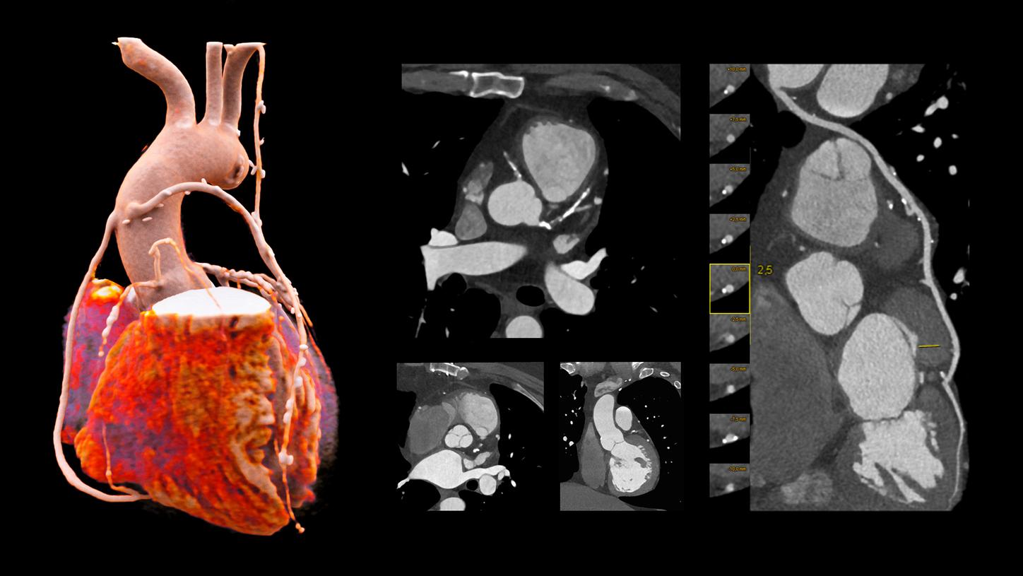
Adaptive Cardio Spiral
120 ㎸
CTDIvol: 13.8 mGy
DLP: 302.2 mGy*cm
Exposure time: 10 s
Scan length: 219 mm
Rotation time: 0.3 s
HR: approx. 50 bpm
Multiple bypass assesment
- Robust cardiac imaging with myExam Companion
- Enhanced delineation of soft and hard plaque thanks to ADMIRE iterative reconstruction
- Cinematic VRT
- 0.8 mm MPR used for reconstructions
- Curved MRP of LAD

Courtesy of Clinica Universidad de Navarra, Pamplona, Spain
Adaptive Cardio Spiral
70 kV
CTDIvol: 6 mGy
DLP:91 mGy*cm
Exposure time: 8 s
Scan length: 150 mm
Rotation time: 0.3 s
HR: < 60 bpm
Low-kV coronary CTA
- Low-kV imaging even for adults thanks to the large power reserves of the Vectron® X-ray tube
- Low-kV imaging helps reduce radiation and contrast media dose
- Cinematic VRTs
- Curved MPRs of the main coronaries
- MIP view of RCA and LAD

Courtesy of Clinica Universidad de Navarra, Pamplona, Spain
Calcium Scoring
90 kV
CTDIvol: 2.23 mGy
DLP: 31mGy*cm
Adaptive Cardio Spiral
120 kV
CTDIvol: 35.5 mGy
DLP: 572 mGy*cm
Exposure time: 7 s
Scan length: 165 mm
Rotation time: 0.3 s
HR: approx. 63 bpm
Robust cardiac imaging for irregular HR
- Restrospective acquisition with Cardio BestPhase for optimal phase reconstruction
- Cinematic VRTs
- Automated curved MPR for the main coronaries and Calsium Scoring thanks to Recon&GO

Courtesy of Clinica Universidad de Navarra, Pamplona, Spain
COVID-19 Assesment
10 kV
Scan time: 2 s
Scan length: 311 mm
Pitch: 1.2
Rotation time: 0.3 s
CTDIvol: 5.15 mGy
DLP: 156 mGy*cm
- High scan speed makes the exam easier for patients who cannot cope well with long breath-hold times
- Cinematic VRT slabs
- 1 mm MPRs in different orientations

Courtesy of Clinica Universidad de Navarra, Pamplona, Spain
CARE Child
70 kV
CTDIvol: 2.03 mGy
DLP: 52 mGy*cm
Exposure time: 1.08 s
Scan length: 220 mm
Pediatric imaging
- 11-year-old paitient
- Optimize dose distribution and special modulation curves through CARE Dose4D
- 1 mm MPRs

Courtesy of University Hospital Erlangen, Erlangen, Germany
Tin Filter
Sn140 kV
CTDIvol: 0.24 mGy
DLP: 8 mGy*cm
Scan length: 325 mm
exposure time: 3.1 s
Low-dose lung examination
- Follow-up of a solitary pulmonary micronodule
- Exceptionally low dose levels, comparable to conventional X-ray, with Tin Filter
- 22 mm MIPs
- 1 mm axial MPR

Courtesy of Clinica Universidad de Navarra,
TwinBeam Dual Energy
AuSn120 kV
CTDIvol: 5.52 mGy
DLP: 188 mGy*cm
Exposure time: 9.8 s
Scan length: 356 mm
Rotation time: 0.3 s
Pitch: 0.3
Spectral CT for assesment of pulmonary embolism
- Automatic generation of iodine maps and Monoenergetic reconstructions
- Monoienergetic reconstruction at 40keV for enhanved iodine contrast, axial MPR 3 mm and MIP 50 mm
- Cinematric VRT

Courtesy of University Hospital Erlangen, Erlangen, Germany
Tin Filter
Sn100 kV
CTDIvol: 0.43 mGy
DLP: 4 mGy*cm
Exposure time: 5.7 s
Scan length: 109 mm
Ultra-low-dose sinus examination
- 2 mm MPRs coronal and axial
- Sinusitis paitient, preoperative study
- Exceptionally low dose levels, comparable to conventional X-ray, with Tin Filter

Courtesy of University Hospital Zurich, Zurich, Swizerland
Tin Filter
Sn130 kV
CTDIvol: 25.48 mGy
DLP: 22 mGy*cm
Exposure time: 6 s
Rotation time: 1 s
Pitch: 0.85
High-resolution inner ear with no dose panalties
- Improved spatial resolution thanks to the combination of:
- 0.6 x 0.7 (IEC) flying focal apot unique with the Vectron® X-ray tube
- Increased detector density of the StellarInfinity detector with 840 channels per row
- 0.6 mm MPR bilateral, axial and coronal reconstruction

Courtesy of University Hospital Zurich, Zurich, Swizerland
Native head
120 kV
CTDIvol: 40.07 mGy
DLP: 732 mGy*cm
Perfusion
70 kV
CTDIvol: 158.11 mGy
DLP: 1,887 mGy*cm
Rotation time: 0.3 s
4D Imaging beyond the detector width
- Entire supratentorial brain coverage with Flex 4D Spiral
- Low-dose techniques such as 70 kV available thanks to the large power reserves of the Vectron® X-ray tube
- Assess perfusion results including penumbra quantification with Auto-Stroke directly at the scanner console

Courtesy of University Hospital Zurich, Zurich, Swizerland
Carotid CTA
90 kV
CTDIvol: 7.07 mGy
DLP: 282 mGy*cm
Rotation time: 0.3 s
Scan length: 40 cm
Surpra-aortic CTA for clot assessment
- The high power reserves of the Vectron® X-ray tube allow low-kV imaging with optimal image quality even in high-attenuation areas like the shoulders
- Low-kV imaging helps resuce radiation and contrast media dose
- Bone-removed MIP automatically reconstructed with Recon&GO
- Curved MPR of left internal carotid, 1 mm

Corutesy of University Hospital Zurich, Zurich, Swizerland
Spectral CT examnation
TwinSpiral
80 kV / Sn150
CTDIvol: 37 mGy
DLP: 659 mGy*cm
Exposure time: 35 s
Scan length: 186 mm
Rule-out of actute bleeding after mechanical thrombectomy
- TwinSpiral supports assessing the presence of iodine deposits, not present in the virtual noncontrast
- Coronal MPR of a follow-up examination demonstrating that the hyperdense areas wer correctly associated to iodine deposits after intervention

Courtesy of University Hospital Erlangen, Erlangen, Germany
TwinSpiral Dual Energy
90 kV / Sn150 kV
CTDIvol: 16.65 mGy
DLP: 417 mGy*cm
Exposure time: 12 s
Pitch: 0.6 (each)
Rotation time: 0.5 s (each)
Spectral assesment of bone marrow
- TwinSpiral Dual Energy helps distingush blood bruises within the bone marrow
- By filtering out the high energies with Tin Filter, TwinSpiral Dual Energy provides optimal spectral separation in bone marrow and to dose penalty

Courtesy of University Hospital Erlangen, Erlangen, Germany
Arterial
100 kV
CTDIvol: 27.7 mGy
DLP: 1,450 mGy*cm
Exposure time: 15.71 s
Scan length: 533 mm
Rotation time 0.5 秒
Bariatric case
- The large power reserves of the Vectron® X-ray tube and the 82 cm bore make imaging possible for the most challenging cases
- HD field of view extends reconstructions up to 82 cm
- Cinematic VRT
- 8 mm MIP
Courtesy of University Hospital Erlangen, Erlangen, Germany
TwinBeam Dual Energy
AuSn120 kV
CTDIvol: 8.06 mGy
DLP: 547 mGy*cm
Exposure time: 18 s
Scan length: 689 mm
Rotation time: 0.3 s
Pitch: 0.3
Full body assesment with apectral CT
- Rich quantitative information with no dose penalty thanks to TwinBeam Dual Energy
- Go beyond HU and quantify ionine uptake in one phase
- Reduce metal artifacts with iMAR (iterative Metal Artifact Reduction), a proven high-end iterative algorithm
Courtesy of University Hospital Erlangen, Erlangen, Germany
Adaptive Cardio Spiral
120 ㎸
CTDIvol: 13.8 mGy
DLP: 302.2 mGy*cm
Exposure time: 10 s
Scan length: 219 mm
Rotation time: 0.3 s
HR: approx. 50 bpm
Multiple bypass assesment
- Robust cardiac imaging with myExam Companion
- Enhanced delineation of soft and hard plaque thanks to ADMIRE iterative reconstruction
- Cinematic VRT
- 0.8 mm MPR used for reconstructions
- Curved MRP of LAD
Courtesy of Clinica Universidad de Navarra, Pamplona, Spain
Adaptive Cardio Spiral
70 kV
CTDIvol: 6 mGy
DLP:91 mGy*cm
Exposure time: 8 s
Scan length: 150 mm
Rotation time: 0.3 s
HR: < 60 bpm
Low-kV coronary CTA
- Low-kV imaging even for adults thanks to the large power reserves of the Vectron® X-ray tube
- Low-kV imaging helps reduce radiation and contrast media dose
- Cinematic VRTs
- Curved MPRs of the main coronaries
- MIP view of RCA and LAD
Courtesy of Clinica Universidad de Navarra, Pamplona, Spain
Calcium Scoring
90 kV
CTDIvol: 2.23 mGy
DLP: 31mGy*cm
Adaptive Cardio Spiral
120 kV
CTDIvol: 35.5 mGy
DLP: 572 mGy*cm
Exposure time: 7 s
Scan length: 165 mm
Rotation time: 0.3 s
HR: approx. 63 bpm
Robust cardiac imaging for irregular HR
- Restrospective acquisition with Cardio BestPhase for optimal phase reconstruction
- Cinematic VRTs
- Automated curved MPR for the main coronaries and Calsium Scoring thanks to Recon&GO
Courtesy of Clinica Universidad de Navarra, Pamplona, Spain
COVID-19 Assesment
10 kV
Scan time: 2 s
Scan length: 311 mm
Pitch: 1.2
Rotation time: 0.3 s
CTDIvol: 5.15 mGy
DLP: 156 mGy*cm
- High scan speed makes the exam easier for patients who cannot cope well with long breath-hold times
- Cinematic VRT slabs
- 1 mm MPRs in different orientations
Courtesy of Clinica Universidad de Navarra, Pamplona, Spain
CARE Child
70 kV
CTDIvol: 2.03 mGy
DLP: 52 mGy*cm
Exposure time: 1.08 s
Scan length: 220 mm
Pediatric imaging
- 11-year-old paitient
- Optimize dose distribution and special modulation curves through CARE Dose4D
- 1 mm MPRs
Courtesy of University Hospital Erlangen, Erlangen, Germany
Tin Filter
Sn140 kV
CTDIvol: 0.24 mGy
DLP: 8 mGy*cm
Scan length: 325 mm
exposure time: 3.1 s
Low-dose lung examination
- Follow-up of a solitary pulmonary micronodule
- Exceptionally low dose levels, comparable to conventional X-ray, with Tin Filter
- 22 mm MIPs
- 1 mm axial MPR
Courtesy of Clinica Universidad de Navarra,
TwinBeam Dual Energy
AuSn120 kV
CTDIvol: 5.52 mGy
DLP: 188 mGy*cm
Exposure time: 9.8 s
Scan length: 356 mm
Rotation time: 0.3 s
Pitch: 0.3
Spectral CT for assesment of pulmonary embolism
- Automatic generation of iodine maps and Monoenergetic reconstructions
- Monoienergetic reconstruction at 40keV for enhanved iodine contrast, axial MPR 3 mm and MIP 50 mm
- Cinematric VRT
Courtesy of University Hospital Erlangen, Erlangen, Germany
Tin Filter
Sn100 kV
CTDIvol: 0.43 mGy
DLP: 4 mGy*cm
Exposure time: 5.7 s
Scan length: 109 mm
Ultra-low-dose sinus examination
- 2 mm MPRs coronal and axial
- Sinusitis paitient, preoperative study
- Exceptionally low dose levels, comparable to conventional X-ray, with Tin Filter
Courtesy of University Hospital Zurich, Zurich, Swizerland
Tin Filter
Sn130 kV
CTDIvol: 25.48 mGy
DLP: 22 mGy*cm
Exposure time: 6 s
Rotation time: 1 s
Pitch: 0.85
High-resolution inner ear with no dose panalties
- Improved spatial resolution thanks to the combination of:
- 0.6 x 0.7 (IEC) flying focal apot unique with the Vectron® X-ray tube
- Increased detector density of the StellarInfinity detector with 840 channels per row
- 0.6 mm MPR bilateral, axial and coronal reconstruction
Courtesy of University Hospital Zurich, Zurich, Swizerland
Native head
120 kV
CTDIvol: 40.07 mGy
DLP: 732 mGy*cm
Perfusion
70 kV
CTDIvol: 158.11 mGy
DLP: 1,887 mGy*cm
Rotation time: 0.3 s
4D Imaging beyond the detector width
- Entire supratentorial brain coverage with Flex 4D Spiral
- Low-dose techniques such as 70 kV available thanks to the large power reserves of the Vectron® X-ray tube
- Assess perfusion results including penumbra quantification with Auto-Stroke directly at the scanner console
Courtesy of University Hospital Zurich, Zurich, Swizerland
Carotid CTA
90 kV
CTDIvol: 7.07 mGy
DLP: 282 mGy*cm
Rotation time: 0.3 s
Scan length: 40 cm
Surpra-aortic CTA for clot assessment
- The high power reserves of the Vectron® X-ray tube allow low-kV imaging with optimal image quality even in high-attenuation areas like the shoulders
- Low-kV imaging helps resuce radiation and contrast media dose
- Bone-removed MIP automatically reconstructed with Recon&GO
- Curved MPR of left internal carotid, 1 mm
Corutesy of University Hospital Zurich, Zurich, Swizerland
Spectral CT examnation
TwinSpiral
80 kV / Sn150
CTDIvol: 37 mGy
DLP: 659 mGy*cm
Exposure time: 35 s
Scan length: 186 mm
Rule-out of actute bleeding after mechanical thrombectomy
- TwinSpiral supports assessing the presence of iodine deposits, not present in the virtual noncontrast
- Coronal MPR of a follow-up examination demonstrating that the hyperdense areas wer correctly associated to iodine deposits after intervention
Courtesy of University Hospital Erlangen, Erlangen, Germany
TwinSpiral Dual Energy
90 kV / Sn150 kV
CTDIvol: 16.65 mGy
DLP: 417 mGy*cm
Exposure time: 12 s
Pitch: 0.6 (each)
Rotation time: 0.5 s (each)
Spectral assesment of bone marrow
- TwinSpiral Dual Energy helps distingush blood bruises within the bone marrow
- By filtering out the high energies with Tin Filter, TwinSpiral Dual Energy provides optimal spectral separation in bone marrow and to dose penalty
Courtesy of University Hospital Erlangen, Erlangen, Germany
Arterial
100 kV
CTDIvol: 27.7 mGy
DLP: 1,450 mGy*cm
Exposure time: 15.71 s
Scan length: 533 mm
Rotation time 0.5 秒
Bariatric case
- The large power reserves of the Vectron® X-ray tube and the 82 cm bore make imaging possible for the most challenging cases
- HD field of view extends reconstructions up to 82 cm
- Cinematic VRT
- 8 mm MIP
Courtesy of University Hospital Erlangen, Erlangen, Germany
TwinBeam Dual Energy
AuSn120 kV
CTDIvol: 8.06 mGy
DLP: 547 mGy*cm
Exposure time: 18 s
Scan length: 689 mm
Rotation time: 0.3 s
Pitch: 0.3
Full body assesment with apectral CT
- Rich quantitative information with no dose penalty thanks to TwinBeam Dual Energy
- Go beyond HU and quantify ionine uptake in one phase
- Reduce metal artifacts with iMAR (iterative Metal Artifact Reduction), a proven high-end iterative algorithm
1/15
技術情報
SOMATOM X.cite
 FAST 3D CameraMyExam CompanionVectron® X線管
ビジュアルガイダンス・患者観察カメラ
GO テクノロジーモバイルワークフロー
82 cm のボア径
StellarInfinity® Detector
低管電圧撮影とTin Filter
マルチパーパステーブル
myNeedle Companion
X-ray tube | Vectron® X-ray tube |
Detector | StellarInfinity detector ® |
Number of acquired slices | 128 |
Rotation time | 0.3 s* |
In-plane temporal resolution | 150 ms* |
Generator power | 105 kW |
Max. mA (70, 80kV) | 1,200 mA |
Gantry opening | 82 cm |
Gantry cooling | Water / Air |
Table load | 340 kg* |
* オプション
このページの情報はお役に立ちましたか?
ありがとうございます。
具体的なコメントがあればご記入ください . 弊社からの連絡が必要な場合はお問い合わせフォームよりをご利用ください
125 / 125
1
コンピューター断層撮影と高度な治療
製品/機能および/またはサービス(本書に記載されている)は、すべての国および/またはすべてのモダリティで市販されているわけではありません。 将来の利用可能性は保証できません。
本ページに記載されているお客様のコメントは、そのお客様の独自の環境で達成された結果に基づいています。 いわゆる「典型的な」病院はなく、また多くの異なる条件が存在するため(例:病院の規模、ケースミックス、導入するITシステムのレベルなど)、他のお客様が同じ結果を得られるという保証はありません。
ゾマトム X
302AABZX00030000
302AABZX00030000
あなたは医療従事者ですか?
このウェブサイトは医療従事者向けのコンテンツを含んでおります。閲覧は医療従事者と限定させていただきます。
