prostate cancer
Cancer awareness

Prostate cancer: Facts, diagnosis, and treatment

Prostate cancer affects many men, yet its early signs can be subtle. Learn more about the risk factors, symptoms, diagnosis, and treatment options.

For those affected by cancer, sometimes it's the little things that can have a big impact on their quality of life. Shelby Moneer of ZERO Prostate Cancer, a non-profit organization for patient advocacy, explains why it's important for patients to share their stories.

Facts

Prostate cancer remains a uniquely challenging and complex illness, with significant complications and unpredictable treatment outcomes. Despite the advances made in recent years, it remains the fifth leading cause of death worldwide and the second most frequent cancer diagnosis in men [1].

Today prostate cancer is already a major cause of death and disability, and the most common form of male cancer in more than 100 countries. With populations aging and life expectancy rising globally, a new analysis forecasts a dramatic surge in cases and deaths over the next 15 years. Diagnoses are projected to increase from 1.4 million a year in 2020 to 2.9 million by 2040 [5].

There are several steps a man can take to potentially reduce the risk of developing prostate cancer. Regular physical activity and a balanced diet can help maintain a healthy weight. And since obesity is linked to an increased risk of aggressive prostate cancer, maintaining a healthy weight can reduce this risk [8].

Men should undergo regular medical check-ups such as:

  • Blood test: Regular testing for prostate cancer can help detect the disease early when it is most treatable.
  • Digital Rectal Exam (DRE): During a digital rectal exam, the doctor feels the back wall of the prostate gland for enlargement, tenderness, lumps or hard spots.

Prostate cancer symptoms can vary and may not be noticeable in the early stages. Initially, a delay in starting urination might be noticed. Waking up multiple times during the night with a strong urge to urinate can disrupt sleep and lead to morning fatigue.
Over time, a burning sensation while urinating might develop. If blood is later observed in the urine and subsequently in the semen, it is crucial to seek medical attention promptly, as this can indicate a serious condition [9].

The image illustrates common symptoms associated with prostate cancer

Diagnosis and Staging

What if a doctor suspects a patient might have prostate cancer?

During diagnosis, a patient will be asked about any symptoms as described above and about possible risk factors including his family history. In case of any suspicion, the doctor might order some tests.

PSA blood test 

Prostate-specific antigen (PSA) is a protein made by normal and malignant cells of the prostate gland. PSA circulates through the body in two ways: either bound to other proteins or freely, not bound. PSA is detected in the blood of males with a normal prostate and is also found in benign (prostatitis, BPH) and malignant prostate tissue [10,11]. 

The PSA test is indicated for the measurement of total PSA (both free and complexed PSA) in conjunction with Digital Rectal Exam (DRE) as an aid in the detection of prostate cancer in men aged 50 years and older. The PSA test is also used as an aid in monitoring patients with prostate cancer. The free PSA (fPSA) test is used in conjunction with the total PSA test in men aged 50 years and older to determine the percent fPSA value. The percent fPSA value can be used as an aid in discriminating between prostate cancer and benign prostatic disease. Prostate cancer risk increases as the fPSA-to-total-PSA ratio decreases [10,12].

Imaging tests for prostate cancer 

Imaging tests play a key role in the diagnosis and staging of prostate cancer. These tests use magnetic fields (magnetic resonance imaging), sound waves (ultrasound), X-rays (computed tomography), or radioactive substances (positron emission tomography) to create pictures of the prostate. One or more imaging tests like the following might be used to look for cancer in the prostate or to help the doctor see the prostate during certain procedures [13].

Magnetic resonance imaging

Magnetic resonance imaging (MRI) is an important tool in managing prostate cancer, as it can help in earlier detection, more precise diagnosis, and treatments that are tailored to each patient. MRI is known for its ability to clearly depict soft tissues and tumors and is especially useful for prostate cancer. Unlike other imaging modalities, MRI uses magnets to create detailed images of the prostate and surrounding tissues. MRI gives doctors information about how likely it is the patient has cancer in the prostate, and it can help them decide if a biopsy is needed and from which parts of the prostate to take samples [13].

If the results of a PSA blood test or other tests suggest the presence of prostate cancer and the suspicion is confirmed on the clinical images, the patient will most likely need prostate biopsy. A core needle biopsy is the main method used to diagnose prostate cancer. It is usually done by an urologist. If cancerous cells are found during a biopsy, they may also be tested for specific gene or protein changes that could influence treatment options [2,15].

  • Transrectal ultrasound (TRUS)-guided systematic biopsy
    A transrectal ultrasound (TRUS)-guided biopsy uses ultrasound to guide a needle in extracting tissue samples from 12 sites in the prostate, primarily targeting the posterior area.
  • Transperineal biopsy (TPB)
    A transperineal biopsy (TPB) uses a template grid to guide needle sampling of the prostate through the perineum, increasing accuracy by covering more areas. In around 26-36% of patients who had TPB before or after TRUS biopsy, new cancerous lesions or more advanced disease were found [16,17].
  • MRI-guided prostate biopsy
    This newer method combines MRI and ultrasound images to target specific areas for biopsy rather than using a blind systematic approach. 
  • Transurethral biopsy
    The transurethral biopsy is a less common technique where a cystoscope is inserted through the urethra to collect prostate tissue samples from the urethral wall.
  • Biopsy of the lymph nodes
    Lymph node biopsy (lymphadenectomy) involves removing one or more nodes to check for cancer. It’s rarely used for prostate cancer but helps assess if the disease has spread to nearby nodes.


Clinical staging helps determine if additional tests are needed. Pathological staging – based on information obtained during surgery, including the removal of the prostate and possibly lymph nodes – is used to determine the Gleason score and grade group.

Determining the stage of prostate cancer is crucial for guiding treatment decisions.
It helps to:

  • assess the risk of cancer recurrence or spread after initial treatment
  • predict the prognosis
  • evaluate the effectiveness of a treatment

The Gleason score grades prostate cancer aggressiveness based on cell appearance in a biopsy.

Treatment

Catching prostate cancer early makes it highly curable.

When treated before it spreads, many people go on to enjoy long, cancer-free lives. There are several therapy options for patients with prostate cancer because the treatment is as individual as the patient who needs it [20,21].
98% five-year survival all stages combined

Watchful waiting and active surveillance (AS) are approaches for men without symptoms or those with other medical conditions. Watchful waiting involves monitoring the patient without treatment until symptoms appear or change. AS also involves monitoring but includes regular tests to detect early signs of cancer progression. Treatment is started if the cancer shows signs of growth.

Patients in good health whose tumor is in the prostate gland only may be treated with surgery to remove the tumor. Different types of surgery are used: 

  • Radical prostatectomy is a surgical procedure to remove the prostate, surrounding tissue, and seminal vesicles. A removal of nearby lymph nodes may be done at the same time. 
  • Pelvic lymphadenectomy is a surgical procedure to remove the lymph nodes in the pelvis. A pathologist views the tissue under a microscope to look for cancer cells. If the lymph nodes contain cancer, the doctor will not remove the prostate and may recommend other treatment. 
  • Transurethral resection of the prostate is a procedure to remove tissue from the prostate using a resectoscope inserted through the urethra. This procedure is sometimes done to relieve symptoms caused by a tumor before other cancer treatment is given.

A resectoscope is a medical instrument used to examine the rectum and lower colon. It consists of a rigid tube with a camera, allowing for diagnosis or minor procedures.

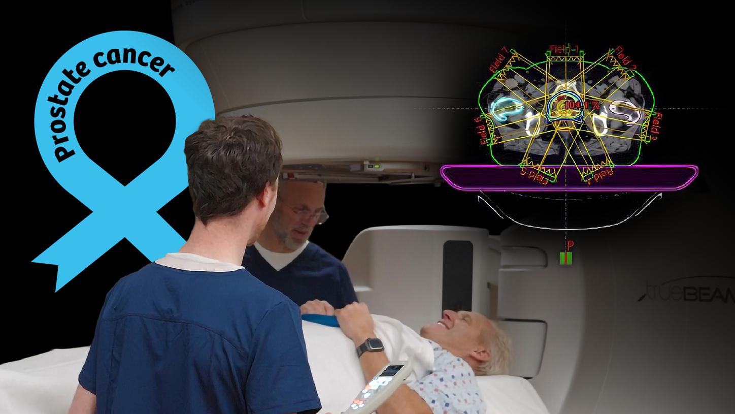
Image of Varian Eclipse Intensity Modulated Radiotherapy plan for prostate cancer patient
IMRT is advanced radiation therapy that targets tumors with adjustable beams, minimizing damage to healthy tissue and reducing side effects.

Radiation therapy (RT) is a cancer treatment that uses high-energy X-rays or other types of radiation to kill cancer cells or keep them from growing. There are different types of radiation therapy: 

External beam therapy, the most common form of RT, is using high-energy X-rays to target and treat disease. The sophisticated machine that delivers radiation therapy is called a linear accelerator, or “linac,” for short. 

Hypofractionated radiation therapy is radiation treatment in which a larger than usual total dose of radiation is given once a day over a shorter period of time compared to standard radiation therapy. It may have increased side effects than standard radiation therapy, depending on the schedules used. 

Brachytherapy implants radioactive sources in the patient to kill cancer cells. In early-stage prostate cancer, the radioactive seeds are placed in the prostate using needles. The placement of the radioactive seeds in the prostate is guided by images from transrectal ultrasound or computed tomography. 

Magnetic resonance imaging can add additional insights for precise planning of prostate radiation therapy. While the prostate is the primary target for the radiation and should receive full dose, it is surrounded by organs sensitive to the effect of radiation, which should be spared as much as possible. MR images can depict this anatomy with increased contrast, therefore assisting the planner in achieving a personalized plan. During the course of treatment, diffusion-weighted MRI can give an indication whether the treatment is working. 

Proton therapy is a form of external beam therapy that uses protons instead of high-energy X-rays. That makes it possible to treat certain types of cancer more precisely and with potentially fewer side effects than is possible with conventional radiation therapy [22]. With proton therapy, the risk of damage to healthy tissues and potential side effects is reduced because a proton beam deposits dose within the tumor site rather than passing all the way through the patient. 

A radiation oncologist will determine the most appropriate treatment for every patient, and the medical team will tailor treatment according to a patient‘s particular situation.


Diffusion-weighted MRI uses specific sequences that measure the diffusion of water molecules to generate contrast in MR images.

Hormone therapy targets cancer by either removing or blocking hormones that fuel its growth. Since hormones – substances produced by glands and circulated in the bloodstream – can drive prostate cancer, this treatment aims to reduce or block male sex hormones that promote tumor growth. This can be achieved through medications, surgery, or other hormonal interventions.
Chemotherapy is a cancer treatment that uses drugs to stop the growth of cancer cells, either by killing the cells or by stopping them from dividing. When chemotherapy is taken by mouth or injected into a vein or muscle, the drugs enter the bloodstream and can reach cancer cells throughout the body.
Targeted therapy is a type of treatment that uses drugs or other substances to identify and attack specific cancer cells. It usually causes less harm to healthy cells than chemotherapy or radiation therapy do.
Immunotherapy is a treatment that uses the patient’s immune system to fight cancer. Substances made by the body or made in a laboratory are used to boost, direct, or restore the body’s natural defenses against cancer. This cancer treatment is a type of biologic therapy.

Theranostics revolutionizes personalized medicine by using molecular imaging and radioisotopes to target the disease. Managing prostate cancer with theranostics advances personalized medicine by diagnosing and treating the disease with individualized precision [23].

During theranostics, doctors first use a radioactive drug to locate and tag cancer cells. These radiotracers, when administered to the patient, bind to specific molecules or cells associated with the disease. Once the tracers identify the disease, therapeutic agents – often also radioactive – are administered. These therapeutic agents are designed to specifically target and destroy the identified diseased cells or tissues. Both the diagnostic and therapeutic drugs are aimed at the same molecular target on the cancer cells, allowing for a precise attack that can enhance treatment outcomes and minimize side effects.

Two images: one pre and one post treatment PSMA PET/CT

Ensuring that prostate cancer is truly gone after treatment involves a vigilant and ongoing process of monitoring and care. The cornerstone of this process is regular PSA testing, especially in the critical first few years post-treatment. Alongside PSA tests, doctors often rely on physical exams, such as digital rectal exams, and imaging techniques like MRI, CT or PSMA PET/CT to check for any signs that cancer might still be present. 

In cases where there’s any uncertainty, a biopsy may be conducted to confirm whether cancer cells remain. Monitoring for new symptoms is also crucial: unexplained bone pain or urinary issues, for instance, could be indicators that need further investigation. In some situations, additional treatments such as hormone therapy or radiation may be recommended to eliminate any residual cancer cells that may have been missed [24-26].

Cancer stage at time of diagnosis: 10-20% of men are diagnosed with metastasized PCa and 80-90% of men are diagnosed with localized PCa

The progression of prostate cancer is often long and slow. Continuous monitoring throughout the patient’s entire journey can help clinicians identify needs and provide supportive interventions to improve overall quality of life [25].

Whole-body MRI incorporating diffusion sequences can improve disease detection in prostate cancer patients with high metastatic bone disease prevalence. syngo.MR OncoTrend enables accurate monitoring of oncological lesions, helping determine whether treatment is effective or needs to potentially be reconsidered. 

Integrating palliative care early may help improve overall quality of life by monitoring symptoms, pain, and stress related to the disease [25,26].

Over the last 30 years, the incidence of prostate cancer has been rising regardless of income level. The number of deaths from prostate cancer worldwide is predicted to rise by 85 percent over the 20-year-period, from 375,000 in 2020 to almost 700,000 by 2040. Experts believe the actual death toll is likely higher due to underdiagnosis and missing data in low and middle-income countries [4]. Higher income countries experienced highest cancer incidence but also the highest decrease in the mortality rates, while lower income countries faced the opposite challenges with lower increase but higher mortality rates [27]. 

In areas with solid testing programs, PSA tests are frequently handled by primary care doctors, often during routine wellness visits. Routine screening can lead to the early detection of prostate cancer, often before symptoms arise. This allows for earlier intervention, which may improve outcomes and reduce mortality in some men. 

While disparities in prostate cancer care still exist, progress is being made. A recent analysis by the American Cancer Society indicates that the overall cancer mortality gap is closing, largely due to a notable decrease in lung and prostate cancer rates. The data highlights a promising outlook, suggesting that advancements in screening, access to care, delivery methods, and clinical trial participation could enhance outcomes for all prostate cancer patients [28].